

doi: 10.1007/s13193-014-0305-8.
Epub 2014 Apr 19.
p53: Revealing the Unusual Suspect: a Study and Field Cancerization Minireview
Affiliations
- PMID: 25114469
- PMCID: PMC4116548
- DOI: 10.1007/s13193-014-0305-8
Item in Clipboard
p53: Revealing the Unusual Suspect: a Study and Field Cancerization Minireview
Indian J Surg Oncol.
2014 Jun.
No abstract available
Figures
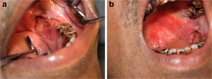
a Photograph showing ulcero-proliferative growth in the left mandibular posterior alveolar region. b Photograph showing erythematous plaque on the hard palate separated from the ulcero-proliferative growth by normal appearing mucosa
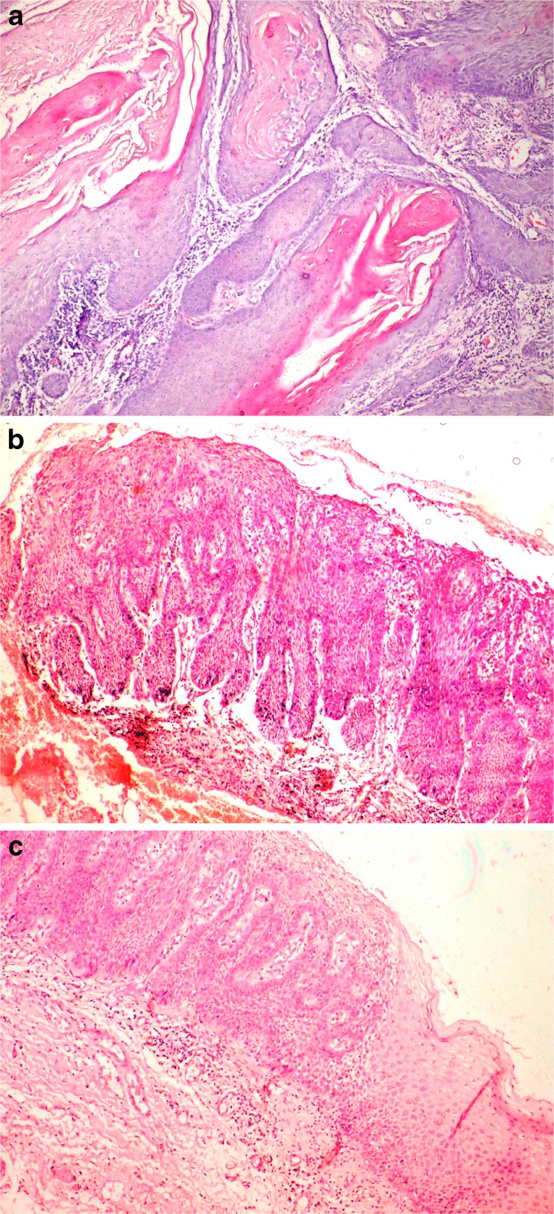
a Photomicrograph (H&E stain, 200×) depicting histopathological features of well differentiated squamous cell carcinoma. b Photomicrograph (H&E stain, 200×) exhibiting histopathological features of moderate to severe dysplasia. c Photomicrograph (H&E stain 200×) showing histologically normal oral mucosa

a Photomicrograph (p53 IHC stain, 400×) revealing intense p53 positivity in all the tumor cells. b Photomicrograph (p53 IHC stain, 400×) showing intense p53 positivity in all cell layers of dysplastic epithelium. c Photomicrograph (p53 IHC stain, 400×) showing intense p53 positivity in basal and suprabasal cell layers of normal epithelium
References
-
- Cruz IB, Snijders PJF, Meijer CJ, Braakhuis BJ, Snow GB, et al. p53 expression above the basal cell layer in oral mucosa is an early event of malignant transformation and has predictive value for developing oral squamous cell carcinoma. J Pathol. 1998;184:360–368. doi: 10.1002/(SICI)1096-9896(199804)184:4<360::AID-PATH1263>3.0.CO;2-H. - DOI - PubMed
-
- Bedi GC, Westra WH, Gabrielson E, Koch W, Sidransky D. Multiple head and neck tumors: evidence for a common clonal origin. Cancer Res. 1996;56:2484–2487. - PubMed
LinkOut - more resources
Full Text Sources
Other Literature Sources
Research Materials
Miscellaneous