. 2014 Jul 29:9:3509-26.
doi: 10.2147/IJN.S63717. eCollection 2014.

Self-assembled nanoparticles based on the c(RGDfk) peptide for the delivery of siRNA targeting the VEGFR2 gene for tumor therapy

Affiliations

Self-assembled nanoparticles based on the c(RGDfk) peptide for the delivery of siRNA targeting the VEGFR2 gene for tumor therapy

Li Liu et al. Int J Nanomedicine. .

Abstract

The clinical application of small interfering RNA (siRNA) has been restricted by their poor intracellular uptake, low serum stability, and inability to target specific cells. During the last several decades, a great deal of effort has been devoted to exploring materials for siRNA delivery. In this study, biodegradable, tumor-targeted, self-assembled peptide nanoparticles consisting of cyclo(Arg-Gly-Asp-d-Phe-Lys)-8-amino-3,6-dioxaoctanoic acid-β-maleimidopropionic acid (hereafter referred to as RPM) were found to be an effective siRNA carrier both in vitro and in vivo. The nanoparticles were characterized based on transmission electron microscopy, circular dichroism spectra, and dynamic light scattering. In vitro analyses showed that the RPM/VEGFR2-siRNA exhibited negligible cytotoxicity and induced effective gene silencing. Delivery of the RPM/VEGFR2 (zebrafish)-siRNA into zebrafish embryos resulted in inhibition of neovascularization. Administration of RPM/VEGFR2 (mouse)-siRNA to tumor-bearing nude mice led to a significant inhibition of tumor growth, a marked reduction of vessels, and a down-regulation of VEGFR2 (messenger RNA and protein) in tumor tissue. Furthermore, the levels of IFN-α, IFN-γ, IL-12, and IL-6 in mouse serum, assayed via enzyme-linked immunosorbent assay, did not indicate any immunogenicity of the RPM/VEGFR2 (mouse)-siRNA in vivo. In conclusion, RPM may provide a safe and effective delivery vector for the clinical application of siRNAs in tumor therapy.

Keywords: gene silencing; self-assembly nanoparticles; siRNA delivery; tumor targeting.


Figures

Figure 1
Figure 1
RPM and RPM/siRNA nanoparticle complexes. Notes: (A) The chemical structure of RPM. (B) Schematic for the formation of RPM/siRNA nanoparticle complexes. (a) Hydrogen bonds between the PEG-MAL chains. (b) Hydrogen bonds between the c(RGDfk) peptide rings. The ring is not a plane structure. (c) The mechanism for the formation of RPM/siRNA nanoparticle complexes. Abbreviations: c(RGDfk), cyclo(Arg-Gly-Asp-d-Phe-Lys); MAL, β-maleimidopropionic acid; PEG, 8-amino-3,6-dioxaoctanoic acid; RPM, c(RGDfk)-PEG-MAL; siRNA, small interfering RNA.
Figure 2
Figure 2
Characterizations of RPM nanoparticles and RPM/siRNA complexes. Notes: (A) Size distribution of (a) RPM nanoparticles and (b) RPM/siRNA complexes. Scale bar is 100 nm (insets: transmission electron microscope images). (B) Circular dichroism spectra of (a) RPM nanoparticles and (b) RPM/siRNA complexes. (C) Stability of naked siRNA and RPM/siRNA complexes in serum. Abbreviations: RPM, cyclo(Arg-Gly-Asp-d-Phe-Lys)-8-amino-3,6-dioxaoctanoic acid-β-maleimidopropionic acid; siRNA, small interfering RNA; Mol Ellip, molar ellipticity.
Figure 3
Figure 3
Confocal laser scanning microscopy images of HUVECs that were transfected with different RPM/siRNA complexes. Notes: (A) Confocal laser scanning microscopy images of HUVECs after 6 hours’ incubation of RPM/siRNA complexes which contain different volume ratio between RPM and siRNA. (B) High-definition DIC images of HUVECs when the volume ratio between RPM and siRNA is 1.5:1. Cell nuclei were counterstained with DAPI (blue) and siRNA was labeled with Cy5 (red). Scale bar is 20 μm. Abbreviations: Cy5, indodicarbocyanine-5; DAPI, 4′,6-diamidino-2-phenylindole; DIC, differential interference contrast; HUVECs, human umbilical vein endothelial cells; RPM, cyclo(Arg-Gly-Asp-d-Phe-Lys)-8-amino-3,6-dioxaoctanoic acid-β-maleimidopropionic acid; siRNA, small interfering RNA.
Figure 4
Figure 4
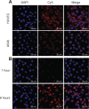
Confocal laser scanning microscopy images of different cell lines and different transfection time in HUVECs. Notes: (A) Different cell lines after incubation with the RPM/siRNA complexes for 6 hours. (B) The cellular uptake and localization of RPM/siRNA complexes in HUVECs at 1 or 6 hours after transfection. Cell nuclei were counterstained with DAPI (blue) and siRNA was labeled with Cy5 (red). Scale bar is 20 μm. Abbreviations: Cy5, indodicarbocyanine-5; DAPI, 4′,6-diamidino-2-phenylindole; HUVEC, human umbilical vein endothelial cell; RPM, cyclo(Arg-Gly-Asp-d-Phe-Lys)-8-amino-3,6-dioxaoctanoic acid-β-maleimidopropionic acid; siRNA, small interfering RNA.
Figure 5
Figure 5
Confocal laser scanning microscopy images of HUVECs that were treated with different ways. Notes: The HUVECs were incubated with anti-integrin αvβ3 antibody for 30 minutes prior to transfection with RPM/siRNA for 6 hours (A) or incubated with the RAPM/siRNA complexes for 6 hours (B). Cell nuclei were counterstained with DAPI (blue) and siRNA was labeled with Cy5 (red). Scale bar is 20 μm. Abbreviations: c(RADfk), cyclo(Arg-Ala-Asp-d-Phe-Lys); c(RGDfk), cyclo(Arg-Gly-Asp-d-Phe-Lys); Cy5, indodicarbocyanine-5; DAPI, 4′,6-diamidino-2-phenylindole; HUVEC, human umbilical vein endothelial cell; MAL, β-maleimidopropionic acid; PEG, 8-amino-3,6-dioxaoctanoic acid; RAPM, c(RADfk)-PEG-MAL; RPM, c(RGDfk)-PEG-MAL; siRNA, small interfering RNA.
Figure 6
Figure 6
Cytotoxicity and gene silencing in vitro. Notes: (A) Cell viability by CCK-8 assay (Beyotime Institute of Biotechnology, Haimen, People’s Republic of China). (B) RT-qPCR analysis of VEGFR2 mRNA levels expressed in HUVECs after a 48-hour transfection (100 nM siRNA:siVEGFR2). All qPCR experiments were done in triplicate and VEGFR2 mRNA expression was normalized to the expression of GAPDH. (C) Western blot analysis of VEGFR2 protein and GAPDH protein expressed in HUVECs after a 72-hour transfection (100 nM siRNA:VEGFR2). Three independent experiments were performed. (D) Quantitative analysis of VEGFR2 protein expression levels. The expression of targeted protein was relative to the expression of GAPDH protein. *P<0.05, **P<0.01, compared with untreated; #P<0.05, ##P<0.01, compared with RPM; &&P<0.01, compared with RPM/control siRNA; $$P<0.01, compared with RPM/siRNA. Lipo2000: Thermo Fisher Scientific (Waltham, MA, USA). Abbreviations: CCK-8, Cell Counting Kit-8; Control siRNA, scramble siRNA; GAPDH, glyceraldehyde-3-phosphate dehydrogenase; HUVECs, human umbilical vein endothelial cells; Lipo2000, Lipofectamine™ 2000; mRNA, messenger RNA; qPCR, quantitative polymerase chain reaction; RPM, cyclo(Arg-Gly-Asp-d-Phe-Lys)-8-amino-3,6-dioxaoctanoic acid-β-maleimidopropionic acid; siRNA, small interfering RNA; VEGFR2, vascular endothelial growth factor receptor 2; RT, reverse transcription.
Figure 7
Figure 7
Antiangiogenic effect of RPM/siVEGFR2 in zebrafish. Notes: (A) After microinjection of RPM/siRNA (siVEGFR2), RPM/control siRNA, naked siRNA (siVEGFR2), ddH2O into the zebrafish embryos which are 10 hours post-fertilization, angiogenesis was imaged using confocal laser scanning microscopy at 72 hours post-fertilization. (B) Relative fluorescence intensity. *P<0.05, **P<0.01, compared with ddH2O; ##P<0.01, compared with naked siRNA (siVEGFR2); &&P<0.01, compared with RPM/control siRNA. Abbreviations: Control siRNA, scramble siRNA; ddH2O, double-distilled water; DLAV, dorsal longitudinal anastomotic vessel; ISVs, intersegmental vessels; RPM, cyclo(Arg-Gly-Asp-d-Phe-Lys)-8-amino-3,6-dioxaoctanoic acid-β-maleimidopropionic acid; siRNA, small interfering RNA; VEGFR2, vascular endothelial growth factor receptor 2.
Figure 8
Figure 8
In vivo localization of nanoparticle complexes with siRNA-Cy5. Notes: (A) In vivo distribution of siRNA-Cy5 in nude mice bearing xenografted luciferase-A549 tumor at the different time points after intravenous injection of RPM/siRNA-Cy5, RAPM/siRNA-Cy5, or naked siRNA-Cy5. Whole-animal bioluminescent imaging was performed to detect luciferase-expressing A549 cells. (B) After 24 hours, ex vivo imaging of tumor and organs excised from BALB/c nude mice. Saline was used as the blank control. Images acquired using IVIS Spectrum imaging system (Xenogen Corporation–Caliper, Ala-meda, CA, USA) with appropriate wavelength (siRNA-Cy5: λex =640 nm, λem =680 nm). All images were scaled to the same minimum and maximum color values. Abbreviations: c(RADfk), cyclo(Arg-Ala-Asp-d-Phe-Lys); c(RGDfk), cyclo(Arg-Gly-Asp-d-Phe-Lys); Cy5, indodicarbocyanine-5; MAL, β-maleimidopropionic acid; PEG, 8-amino-3,6-dioxaoctanoic acid; RAPM, c(RADfk)-PEG-MAL; RPM, c(RGDfk)-PEG-MAL; siRNA, small interfering RNA; siRNA-Cy5, Cy5-labeled siRNA.
Figure 9
Figure 9
Intravenous injection of RPM/siRNA (siVEGFR2) results in reduced tumor growth in mice. Notes: The body weight (A) and the tumor volume (B) were measured every 3 days. Statistical significance compared with the RPM/siRNA group: *P<0.05, **P<0.01. (C) Tumor bioluminescence intensity of each group at day 0 and day 20. Abbreviations: Control siRNA, scramble siRNA; RPM, cyclo(Arg-Gly-Asp-d-Phe-Lys)-8-amino-3,6-dioxaoctanoic acid-β-maleimidopropionic acid; siRNA, small interfering RNA; VEGFR2, vascular endothelial growth factor receptor 2.
Figure 10
Figure 10
Assessment of potential immunostimulatory effects, renal and liver toxicity, and gene silencing of RPM/siRNA (siVEGFR2) complexes in vivo. Notes: (A) Examination of mouse IFN-α, IFN-γ, IL-12, and IL-6 levels in the serum of nude mice bearing xenografted luciferase-A549 tumors at (a) 6 and (b) 24 hours following administration via ELISA test. No differences were found between groups at different time points. (B) Creatinine and ALT levels in the serum. (C) Real-time qPCR analysis of the VEGFR2 mRNA levels expressed in tumors after the last administration. All qPCR experiments were done in triplicate and VEGFR2 mRNA expression was normalized to 18s mRNA. (D) (a) Western blot analysis of VEGFR2 protein and GAPDH protein expressed in tumors after the last administration. (b) Quantitative analysis of VEGFR2 protein expression levels. Data are representative of three independent experiments performed in triplicate, and the expression of targeted protein was relative to the expression of GAPDH protein. (E) Frozen sections of luciferase-A549 tumors were stained with CD31 (brown) at 200× original magnification. **P<0.01, compared with saline; ##P<0.01, compared with RPM/control siRNA. Abbreviations: ALT, alanine aminotransferase; CD31, platelet endothelial cell adhesion molecule-1 (also known as cluster of differentiation 31); Control siRNA, scramble siRNA; ELISA, enzyme-linked immunosorbent assay; GAPDH, glyceraldehyde-3-phosphate dehydrogenase; mRNA, messenger RNA; qPCR, quantitative polymerase chain reaction; RPM, cyclo(Arg-Gly-Asp-d-Phe-Lys)-8-amino-3,6-dioxaoctanoic acid-β-maleimidopropionic acid; siRNA, small interfering RNA; VEGFR2, vascular endothelial growth factor receptor 2.

Similar articles

Cited by

References

    1. Zamore PD, Tuschl T, Sharp PA, Bartel DP. RNAi: double-stranded RNA directs the ATP-dependent cleavage of mRNA at 21 to 23 nucleotide intervals. Cell. 2000;101(1):25–33. - PubMed
    1. de Fougerolles A, Vornlocher HP, Maraganore J, Lieberman J. Interfering with disease: a progress report on siRNA-based therapeutics. Nat Rev Drug Discov. 2007;6:443–453. - PMC - PubMed
    1. Iorns E, Lord CJ, Turner N, Ashworth A. Utilizing RNA interference to enhance cancer drug discovery. Nat Rev Drug Discov. 2007;6:556–568. - PubMed
    1. Aliabadi HM, Landry B, Sun C, Tang T, Uludağ H. Supramolecular assemblies in functional siRNA delivery: where do we stand? Biomaterials. 2012;33:2546–2569. - PubMed
    1. Danhier F, Lecouturier N, Vroman B, et al. Paclitaxel-loaded PEGylated PLGA-based nanoparticles: in vitro and in vivo evaluation. J Control Release. 2009;133:11–17. - PubMed

Publication types

MeSH terms

LinkOut - more resources