Review
. 2014 Jul 28:10:583-95.
doi: 10.2147/TCRM.S48746. eCollection 2014.

Optimal management of renal artery fibromuscular dysplasia

Affiliations
Review

Optimal management of renal artery fibromuscular dysplasia

Anders Gottsäter et al. Ther Clin Risk Manag. .

Abstract

Fibromuscular dysplasia (FMD) is a nonatherosclerotic, noninflammatory angiopathy of unknown cause affecting medium-sized (most commonly renal) arteries and causing renovascular hypertension. The most common medial multifocal type of FMD (with the "string of beads" appearance) is more than four times more prevalent in females than in males. FMD accounts for up to 10% of cases of renovascular hypertension. Compared with patients with atherosclerotic renal artery stenosis, patients with FMD are younger, have fewer risk factors for atherosclerosis, and a lower occurrence of atherosclerosis in other vessels. The etiology is multifactorial, including vessel wall ischemia and smoking, as well as hormonal and genetic factors. Intra-arterial digital subtraction angiography is still the gold standard for exclusion or confirmation of renal artery stenosis caused by FMD, at least in young patients, who more often have lesions in branches of the renal artery. For FMD patients with atherosclerosis and those who are older (>50-55 years), significant renal artery stenosis may be confirmed or excluded with ultrasonography. The FMD lesion is typically truncal or distal, whereas atherosclerotic lesions are more often proximal or ostial. Treatment options are medical, endovascular (percutaneous transluminal renal angioplasty [PTRA]), and surgical. Invasive treatment should be considered when hypertension cannot be controlled with antihypertensive drugs and in patients with impaired renal function or ischemic nephropathy. PTRA has become the treatment of choice and normally yields good results, especially in unifocal disease and young patients. Pressure gradients are normally completely abolished, and there is no indication for stent placement. Surgical revascularization is indicated after PTRA complications; thrombosis, perforation, progressive dissection, repeated PTRA failure or restenosis. Centralization of handling is recommended.

Keywords: arterial hypertension; fibromuscular dysplasia; percutaneous transluminal renal angioplasty; renal artery stenosis; renal artery surgery.


Figures

Figure 1
Figure 1
Typical selective angiographic multifocal appearance of medial fibrodysplasia with “string of beads” appearance in the distal main artery before (A) and after (B) PTA. Note: It is difficult to visualize that the 45 mmHg pressure gradient disappeared and a favorable effect on blood pressure was achieved. Abbreviation: PTA, percutaneous transluminal angioplasty.
Figure 2
Figure 2
Short unifocal stenosis of the distal main artery, perhaps of the perimedial dysplastic type, before (A) and after (B) percutaneous transluminal angioplasty.
Figure 3
Figure 3
Medial fibrodysplasia with dense fibrous connective tissue in the outer layer of the medial layer, disordered inner medial smooth muscle, and collagen deposition. Composite of hematoxylin and elastin staining. Note: (Courtesy of Dr J Malina, Department of Pathology, Skåne University Hospital, Malmö, Sweden).
Figure 4
Figure 4
Magnetic resonance angiography, raising suspicion for bilateral fibromuscular dysplasia. Notes: The right renal artery changes caused significant pressure gradients and were treated with percutaneous transluminal angioplasty. Note the absence of intrarenal branches, that are affected in 25% of patients with fibromuscular dysplasia and are not visualized with magnetic resonance angiography.
Figure 5
Figure 5
Angiography of a pregnant woman (week 16) with uncontrolled hypertension and bruits over the renal arteries. Notes: Angiography with lead aprons covering the lower abdomen and as little radiation and contrast as possible showed bilateral fibromuscular dysplasia (A), arrows, which was treated with percutaneous transluminal angioplasty. Different stages of treatment are shown (BD). The radiation to the fetus was <0.5 mSv, hypertension improved, and the pregnancy was completed successfully. Post partum control showed restenosis that required repeat percutaneous transluminal angioplasty bilaterally (E) with persisting good results thereafter.
Figure 6
Figure 6
Endovascular treatment of renal artery pseudoaneurysm. Notes: Woman with bilateral fibromuscular dysplasia who 15 years earlier had undergone patch angioplasty after failed endovascular treatment (pressure gradient 50 mmHg, recoiling) of the left renal artery, and nephrectomy of an atrophic right kidney. Routine follow-up 15 years later revealed a 3–4 cm pseudoaneurysm around the patch angioplasty (A), which was able to be treated with endovascular placement of a covered stent graft (B,C) and had a good outcome.
Figure 7
Figure 7
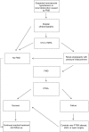
Suggested management principles for fibromuscular dysplasia. Abbreviations: FMD, fibromuscular dysplasia; CTA, computed tomography angiography; MRA, magnetic resonance angiography; PTRA, percutaneous transluminal renal angioplasty.

Similar articles

Cited by

References

    1. ASTRAL investigators Revascularization versus medical therapy for renal-artery stenosis. N Engl J Med. 2009;361(20):1953–1362. - PubMed
    1. Bax L, Mali WP, Buskens E, et al. STAR Study Group The benefit of STent placement and blood pressure and lipid-lowering for the prevention of progression of renal dysfunction caused by Atherosclerotic ostial stenosis of the renal artery. The STAR-study: rationale and study design. J Nephrol. 2003;16(6):807–812. - PubMed
    1. Cooper CJ, Murphy TP, Cutlip DE, et al. CORAL Investigators Stenting and medical therapy for atherosclerotic renal-artery stenosis. N Engl J Med. 2014;370(1):13–22. - PMC - PubMed
    1. Plouin PF, Perdu J, La Batide-Alanore A, et al. Fibromuscular dysplasia. Orphanet J Rare Dis. 2007;2:28. - PMC - PubMed
    1. Andreoni KA, Weeks SM, Gerber DA, et al. Incidence of donor renal fibromuscular dysplasia: does it justify routine renal angiography? Transplantation. 2002;73(7):1112–1116. - PubMed

LinkOut - more resources