Review
. 2013 Apr 30:6:33-49.
doi: 10.4137/MRI.S10640. eCollection 2013.

Magnetic resonance in the detection of breast cancers of different histological types

Affiliations
Review

Magnetic resonance in the detection of breast cancers of different histological types

Rebecca M Mayrhofer et al. Magn Reson Insights. .

Abstract

Breast cancer incidence is increasing worldwide. Early detection is critical for long-term patient survival, as is monitoring responses to chemotherapy for management of the disease. Magnetic resonance imaging and spectroscopy (MRI/MRS) has gained in importance in the last decade for the diagnosis and monitoring of breast cancer therapy. The sensitivity of MRI/MRS for anatomical delineation is very high and the consensus is that MRI is more sensitive in detection than x-ray mammography. Advantages of MRS include delivery of biochemical information about tumor metabolism, which can potentially assist in the staging of cancers and monitoring responses to treatment. The roles of MRS and MRI in screening and monitoring responses to treatment of breast cancer are reviewed here. We rationalize how it is that different histological types of breast cancer are differentially detected and characterized by MR methods.

Keywords: 1H nuclear magnetic resonance spectroscopy; 31P nuclear magnetic resonance spectroscopy; breast cancer; diffusion weighted imaging; dynamic contrast enhanced imaging; magnetic resonance imaging.


Figures
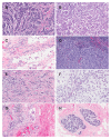
Figure 1
Figure 1
Photomicrographs of various histological types of breast cancers provided by National University Hospital, Singapore. (A) Haematoxylin and eosin (H&E) 40×, invasive ductal carcinoma. Note that the invasive tumor exhibits no features of any special histological type. The tumor is arranged in cords, clusters and trabeculae with scanty tubule formation and several mitoses per high-power image field. (B) H&E 40×, invasive micropapillary carcinoma (a rare variant of invasive ductal carcinoma): the tumor clusters show irregular central spaces, surrounded by artefactually retracted stromal spaces. Some of the clusters have reversed polarity (an “inside out“ morphology). (C) H&E 40×, lobular carcinoma: the tumor shows uniform tumor cells mostly arranged in a single file. Some of the tumor cells show intracytoplasmic luminae. (D) H&E 40×, medullary carcinoma: the tumor is composed of a syncytial sheet of pleomorphic cells with no glandular differentiation. The stroma contains prominent lymphoplasmacytic infiltrate. (E) H&E 40×, tubular carcinoma: the tumor is highlighted by irregularly distributed rounded and angulated tubules with open luminae. The tubules are lined by a single layer of epithelial cells and are surrounded by desmoplastic stroma. (F) H&E 40×, mucinous carcinoma: clusters of uniform tumor cells floating in lakes of mucin. (G) H&E 40×, inflammatory breast cancer: characteristic dermal lymphatic invasion by carcinoma associated with surrounding lymphoplasmacytic inflammatory infiltrate. (H) healthy breast tissue: terminal duct-lobular units surrounded by fibro-myxoid stroma. These are lined by an inner epithelial and an outer myoepithelial cell layer. The full time scale is ~10 min.
Figure 2
Figure 2
Illustration of the different types of kinetic curves from DCE MRI. Notes: Type I is a progressive enhancement pattern that shows a continuous increase in signal intensity over time. The Type II curve has a plateau pattern where there is initial contrast-molecule uptake followed by a plateau phase and the result is suggestive of malignant tumor. The Type III curve is a washout pattern where there is initial rapid uptake followed by reduction in signal enhancement and it is strongly suggestive of malignancy.
Figure 3
Figure 3
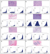
Analyses of the red, blue, and green pixel intensities in the different breast cancer types for the microphotographs provided by National University Hospital, Singapore: (A) invasive ductal carcinoma (IDC). (B) invasive micropapillary carcinoma (a form of IDC). (C) invasive lobular carcinoma (ILC). (D) medullary carcinoma (MC). (E) tubular carcinoma (TC). (F) mucinous carcinoma (MuC). (G) inflammatory breast cancer ‘(IBC). (H) healthy breast tissue. Note: The unique shapes of the histograms for the different colors and breast cancer types.
Figure A3.2
Figure A3.2
Number of papers published per year on breast cancer diagnosis with MRI/MRS. Note: The search engine Scopus was used with the keywords as indicated in the figure.

Similar articles

Cited by

References

    1. Ferlay J, Héry C, Autier P, Sankaranarayanan R. Global burden of breast cancer. Breast Cancer Epidemiol. 2010:1–19.
    1. Parkin DM, Fernández LM. Use of statistics to assess the global burden of breast cancer. Breast J. 2006;12(Suppl 1):S70–80. - PubMed
    1. Ferlay J, Shin HR, Bray F, Forman D, Mathers C, Parkin DM. Estimates of worldwide burden of cancer in 2008: GLOBOCAN 2008. Int J Cancer. 2010;127:2893–917. - PubMed
    1. Leach M, Verrill M, Glaholm J, et al. Measurements of human breast cancer using magnetic resonance spectroscopy: a review of clinical measurements and a report of localized 31P measurements of response to treatment. NMR Biomed. 1998;11(7):314–40. - PubMed
    1. Korkola J, DeVries S, Fridlyand J, et al. Differentiation of lobular versus ductal breast carcinomas by expression microarray analysis. Cancer Res. 2003;63(21):7167–75. - PubMed

LinkOut - more resources