. 2014:2014:961913.
doi: 10.1155/2014/961913. Epub 2014 Jul 9.

Clinical presentation and magnetic resonance findings in sellar tuberculomas

Affiliations

Clinical presentation and magnetic resonance findings in sellar tuberculomas

Dulce Bonifacio-Delgadillo et al. Case Rep Med. 2014.

Abstract

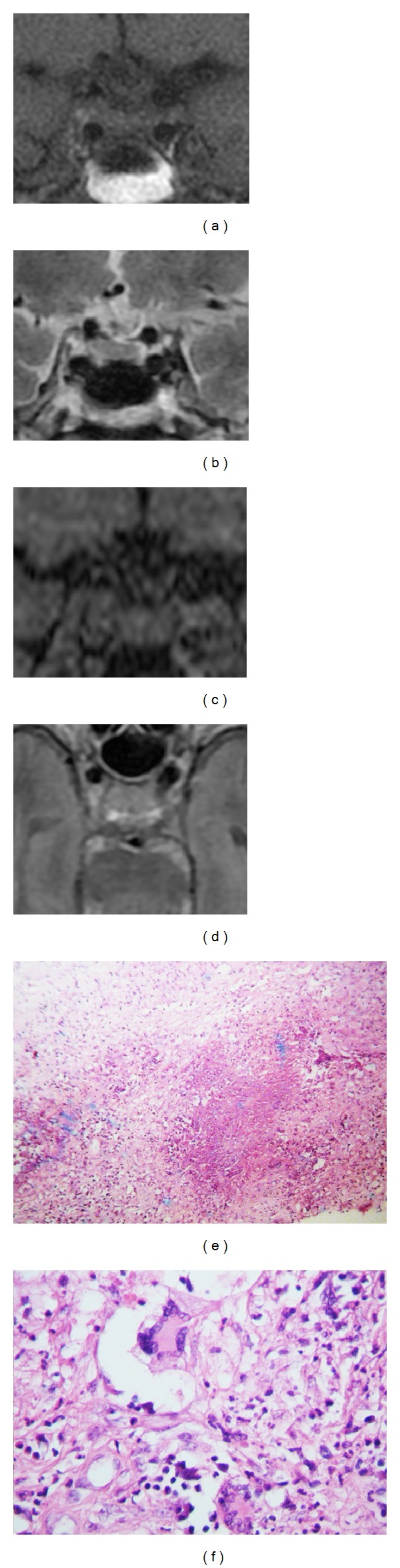
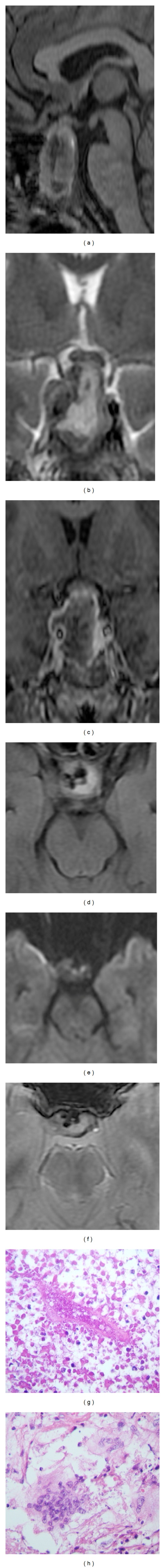
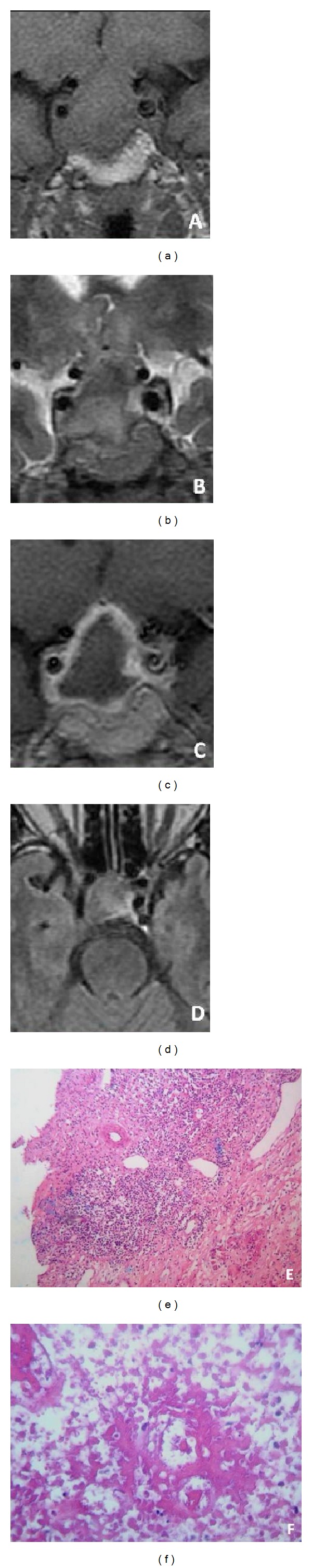
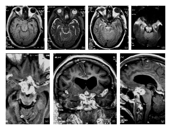
Background and Importance. Sellar tuberculomas are extremely rare lesions with nonspecific clinical manifestations. The tuberculous infection of the pituitary gland and sellar region is characterized by the presence of an acute or chronic inflammatory reaction and may occur in the absence of systemic tuberculosis. The diagnosis is difficult prior to the surgery. An adequate diagnostic and antituberculous drugs usually result in a good outcome. Clinical Presentation. We report four cases of sellar tuberculoma, 3/1 female/male, age range: 50-57 years. All patients had visual disturbances and low levels of cortisol. Conclusion. The clinical diagnosis of sellar tuberculoma is a challenge and should be suspected when a sellar lesion shows abnormal enhancement pattern and stalk involvement, and absence of signal suppression in FLAIR.


Figures

Figure 1
Figure 1
MRI T1-weighted (a), T2-weighted (b), and contrast enhanced T1-weighted (c) of a 67-year-old woman presenting with impaired vision and temporal visual field deficit two months before admission. MRI was done two months after the surgery with pterional approach and showed persistence of intrasellar lesion with stalk involvement. Photomicrograph ×40 H & E (e) and photomicrograph ×400 H & E (f) show central caseous necrosis with lymphocytic inflammatory infiltrate, extensive fibrosis, Langhans multinucleated giant cell with epithelioid cells, and lymphocytes, with few polymorphonuclear.
Figure 2
Figure 2
(a) MRI sagittal T1-weighted, (b) coronal T2-weighted images, (c) coronal T1-weighted images after contrast media administration, (d) axial T2-FLAIR-weighted images, (e) axial diffusion weighted images, and (f) T2∗-weighted images of a 50-year-old woman with headache associated with raised pressure and vertical diplopia revealed a sellar and suprasellar lesion with hemorrhagic areas displacing dorsally the chiasm, thickening the infundibulum and cavernous sinus extension predominantly to the right side. (g) Photomicrograph ×100 H & E and (h) photomicrograph ×400 H & E showed pituitary parenchymal necrosis with blood vessel and group of multinucleated giant cells surrounded by lymphocytes and epithelioid cells.
Figure 3
Figure 3
(a) Coronal T1-weighted images, (b) coronal T2-weighted images, and (c) coronal T1-weighted images after contrast media administration views of initial MRI of a 59-year-old man presented with of fever, nausea, vomiting, polyuria, and loss visual acuity. Intra- and suprasellar lesion contacting bilaterally the gyrus rectus and the chiasm. (e) Photomicrograph ×100 H & E and (f) photomicrograph ×100 H & E showed extensive fibrosis with lymphocytic infiltration and areas of trapped pituitary cells, vasculitis with blood vessel necrosis, adjacent tissue, and few pituitary cells.
Figure 4
Figure 4
(a) Axial T1-weighted images, (b) axial T2-weighted images, and (c) axial T2- and T2-FLAIR-weighted images after contrast media administration. (e) Axial plane, (f) coronal, (g) sagittal MRI images of 57-year-old woman, six years before she had tuberculous meningitis, obstructive hydrocephalus secondary to multiple coalescent nodular well-defined images; the biggest was localized in the sella, the lesion in contact with the chiasm, and extends to cavernous sinuses. Multiple lesions were localized in basal cisterns and both lateral fissures and the retrosellar extension of the lesions cause brain stem compression.

References

    1. von Groote-Bidlingmaier F, Diacon AH. Medical treatment of tuberculosis-update 2011. Therapeutische Umschau Revue Therapeutique . 2011;68(7):395–401. - PubMed
    1. Sunil K, Menon R, Goel N, et al. Pituitary tuberculosis. Journal of Association of Physicians of India. 2007;55:453–456. - PubMed
    1. Arunkumar MJ, Rajshekhar V. Intrasellar tuberculoma presenting as pituitary apoplexy. Neurology India. 2001;49(4):407–410. - PubMed
    1. Garlan HG, Armitage G. Intracranial tuberculoma. The Journal of Pathology and Bacteriology. 1933;37(3):461–471.
    1. Coleman CC, Meredith JM. Diffuse tuberculosis of the pituitary gland simulating tumor, with postoperative recovery. Archives of Neurology and Psychiatry. 1940;44:1076–1085.

LinkOut - more resources