

Multimodality 3-dimensional image integration for congenital cardiac catheterization
- PMID: 25114757
- PMCID: PMC4117323
- DOI: 10.14797/mdcj-10-2-68
Multimodality 3-dimensional image integration for congenital cardiac catheterization
Abstract
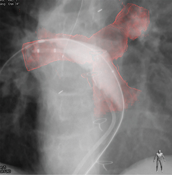
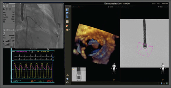
Cardiac catheterization procedures for patients with congenital and structural heart disease are becoming more complex. New imaging strategies involving integration of 3-dimensional images from rotational angiography, magnetic resonance imaging (MRI), computerized tomography (CT), and transesophageal echocardiography (TEE) are employed to facilitate these procedures. We discuss the current use of these new 3D imaging technologies and their advantages and challenges when used to guide complex diagnostic and interventional catheterization procedures in patients with congenital heart disease.
Keywords: 3-dimensional rotational angiography; 3-dimensional transesophageal echocardiography; 3D TEE; 3DRA; CT roadmap; EchoNavigator; HeartNavigator; MRI roadmap; congenital heart disease; rotational angiography; structural heart disease.
Conflict of interest statement
Figures


References
-
- Soderman M, Babic D, Homan R, Andersson T. 3D roadmap in neuroangiography: technique and clinical interest. Neuroradiology. 2005 Oct;47(10):735–40.. - PubMed
-
- Berger MO, Anxionnat R, Kerrien E, Picard L, Söderman M. A methodology for validating a 3D imaging modality for brain AVM delineation: Application to 3DRA. Comput Med Imaging Graph. 2008 Oct;32(7):544–53.. - PubMed
-
- Krishnaswamy A, Tuzcu EM, Kapadia SR. Three-dimensional computed tomography in the cardiac catheterization laboratory. Catheter Cardiovasc Interv. 2011 May 1;77(6):860–5.. - PubMed
Publication types
MeSH terms
LinkOut - more resources
Full Text Sources
Medical
