

Automated tissue classification framework for reproducible chronic wound assessment
- PMID: 25114925
- PMCID: PMC4121018
- DOI: 10.1155/2014/851582
Automated tissue classification framework for reproducible chronic wound assessment
Abstract
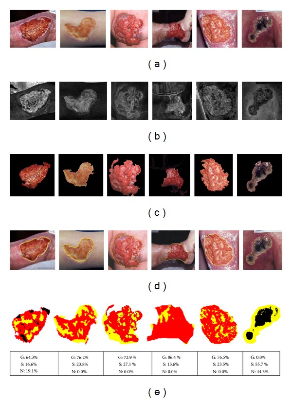
The aim of this paper was to develop a computer assisted tissue classification (granulation, necrotic, and slough) scheme for chronic wound (CW) evaluation using medical image processing and statistical machine learning techniques. The red-green-blue (RGB) wound images grabbed by normal digital camera were first transformed into HSI (hue, saturation, and intensity) color space and subsequently the "S" component of HSI color channels was selected as it provided higher contrast. Wound areas from 6 different types of CW were segmented from whole images using fuzzy divergence based thresholding by minimizing edge ambiguity. A set of color and textural features describing granulation, necrotic, and slough tissues in the segmented wound area were extracted using various mathematical techniques. Finally, statistical learning algorithms, namely, Bayesian classification and support vector machine (SVM), were trained and tested for wound tissue classification in different CW images. The performance of the wound area segmentation protocol was further validated by ground truth images labeled by clinical experts. It was observed that SVM with 3rd order polynomial kernel provided the highest accuracies, that is, 86.94%, 90.47%, and 75.53%, for classifying granulation, slough, and necrotic tissues, respectively. The proposed automated tissue classification technique achieved the highest overall accuracy, that is, 87.61%, with highest kappa statistic value (0.793).
Figures


References
-
- Cutting K, Tong A. Wound Physiology and Moist Wound Healing. Holsworthy, UK: Medical Communications Ltd; 2003.
-
- Gupta N, Gupta SK, Shukla VK, Singh SP. An Indian community-based epidemiological study of wounds. Journal of Wound Care. 2004;13(8):323–325. - PubMed
-
- Romanelli M, Gaggio G, Piaggesi A, Coluccia M, Rizello F. Technological advances in wound bed measurements. Wounds. 2002;14(2):58–66.
-
- Kolesnik M, Fexa A. Multi-dimensional color histograms for segmentation of wounds in images. (Lecture Notes in Computer Science).Image Analysis and Recognition. 2005;3656:1014–1022.
Publication types
MeSH terms
LinkOut - more resources
Full Text Sources
Other Literature Sources
Medical
