

Nitinol-based nanotubular coatings for the modulation of human vascular cell function
- PMID: 25115216
- PMCID: PMC4945101
- DOI: 10.1021/nl501523v
Nitinol-based nanotubular coatings for the modulation of human vascular cell function
Abstract
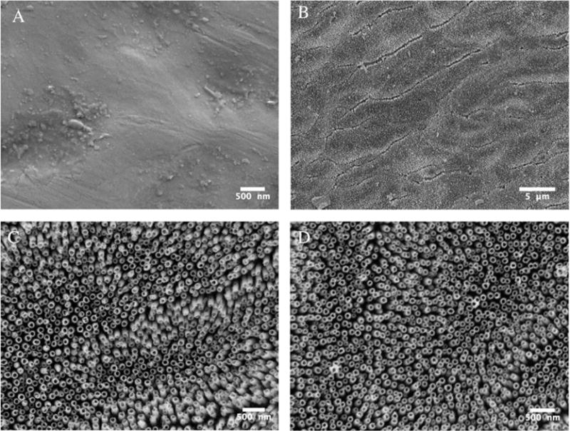
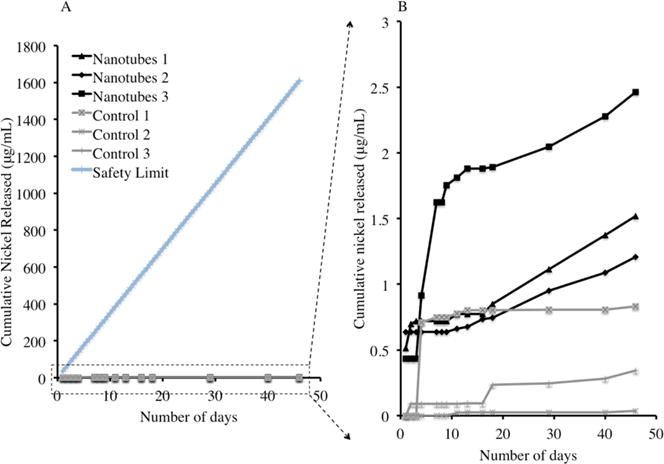
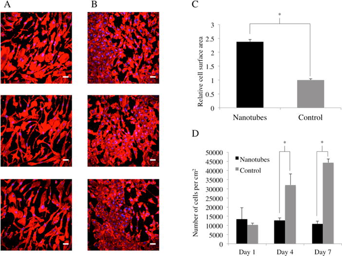
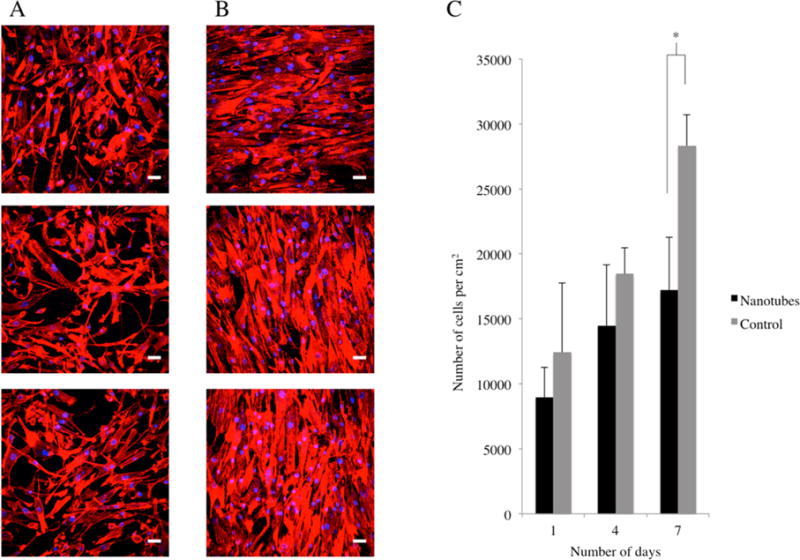
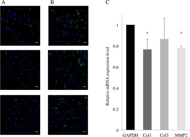
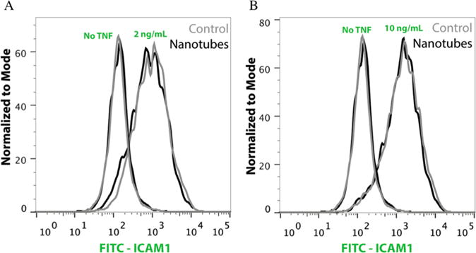
In this study, we describe the synthesis of an upright nanotubular coating with discrete, exposed nanotubes on top of superelastic Nitinol via anodization and characterization of the surface elemental composition and nickel release rates. We demonstrate, for the first time, that this coating could improve re-endothelialization by increasing the cell spreading and migration of primary human aortic endothelial cells on Nitinol. We also show the potential for reducing neointimal hyperplasia by decreasing the proliferation and expression of collagen I and MMP-2 in primary human aortic smooth muscle cells (HASMC). Furthermore, we did not observe the nanotubular surface to induce inflammation through ICAM-1 expression in HASMC as compared to the flat control. This coating could be used to improve Nitinol stents by reducing restenosis rates and, given the extensive use of Nitinol in other implantable devices, act as a generalized coating strategy for other medical devices.
Keywords: Human Aortic Endothelial Cells; Human Aortic Smooth Muscle Cells; Nanotubes; Nitinol; Restenosis; Stents.
Conflict of interest statement
The authors declare no competing financial interest.
Figures


Similar articles
-
Nitinol-Based Nanotubular Arrays with Controlled Diameters Upregulate Human Vascular Cell ECM Production.ACS Biomater Sci Eng. 2016 Mar 14;2(3):409-414. doi: 10.1021/acsbiomaterials.5b00553. Epub 2016 Feb 11. ACS Biomater Sci Eng. 2016. PMID: 27942579 Free PMC article.
-
Long noncoding RNA expression analysis reveals the regulatory effects of nitinol-based nanotubular coatings on human coronary artery endothelial cells.Int J Nanomedicine. 2019 May 7;14:3297-3309. doi: 10.2147/IJN.S204067. eCollection 2019. Int J Nanomedicine. 2019. PMID: 31190794 Free PMC article.
-
Effect of a novel peptide, WKYMVm- and sirolimus-coated stent on re-endothelialization and anti-restenosis.J Mater Sci Mater Med. 2015 Oct;26(10):251. doi: 10.1007/s10856-015-5585-1. Epub 2015 Oct 5. J Mater Sci Mater Med. 2015. PMID: 26438653
-
Mechanisms of smooth muscle cell proliferation and endothelial regeneration after vascular injury and stenting: approach to therapy.Circ J. 2011;75(6):1287-96. doi: 10.1253/circj.cj-11-0366. Epub 2011 Apr 29. Circ J. 2011. PMID: 21532177 Review.
-
Effect of Nitinol surface with nanotubes and/or ordered nanopores on cell behavior.Metallomics. 2022 Feb 23;14(1):mfac002. doi: 10.1093/mtomcs/mfac002. Metallomics. 2022. PMID: 35084501 Review.
Cited by
-
Nitinol-Based Nanotubular Arrays with Controlled Diameters Upregulate Human Vascular Cell ECM Production.ACS Biomater Sci Eng. 2016 Mar 14;2(3):409-414. doi: 10.1021/acsbiomaterials.5b00553. Epub 2016 Feb 11. ACS Biomater Sci Eng. 2016. PMID: 27942579 Free PMC article.
-
Recent advances to accelerate re-endothelialization for vascular stents.J Tissue Eng. 2017 Sep 28;8:2041731417731546. doi: 10.1177/2041731417731546. eCollection 2017 Jan-Dec. J Tissue Eng. 2017. PMID: 28989698 Free PMC article.
-
Advancing Nitinol: From heat treatment to surface functionalization for nickel-titanium (NiTi) instruments in endodontics.Bioact Mater. 2022 Sep 27;22:91-111. doi: 10.1016/j.bioactmat.2022.09.008. eCollection 2023 Apr. Bioact Mater. 2022. PMID: 36203965 Free PMC article.
-
Effectiveness of Direct Laser Interference Patterning and Peptide Immobilization on Endothelial Cell Migration for Cardio-Vascular Applications: An In Vitro Study.Nanomaterials (Basel). 2022 Apr 5;12(7):1217. doi: 10.3390/nano12071217. Nanomaterials (Basel). 2022. PMID: 35407334 Free PMC article.
-
Programming of Regulatory T Cells In Situ for Nerve Regeneration and Long-Term Patency of Vascular Grafts.Research (Wash D C). 2022 Jul 19;2022:9826426. doi: 10.34133/2022/9826426. eCollection 2022. Research (Wash D C). 2022. PMID: 35966759 Free PMC article.
References
-
- Rajagopal V. Coronary Restenosis: A Review of Mechanisms and Management. Am J Med. 2003;115:547–553. - PubMed
-
- Lüscher TF, Steffel J, Eberli FR, Joner M, Nakazawa G, Tanner FC, Virmani R. Drug-Eluting Stent and Coronary Thrombosis: Biological Mechanisms and Clinical Implications. Circulation. 2007;115:1051–1058. - PubMed
-
- Khan W, Farah S, Domb AJ. Drug Eluting Stents: Developments and Current Status. J Controlled Release. 2012:703–712. - PubMed
-
- Mani G, Feldman MD, Patel D, Agrawal CM. Coronary Stents: A Materials Perspective. Biomaterials. 2007;28:1689–1710. - PubMed
Publication types
MeSH terms
Substances
Grants and funding
LinkOut - more resources
Full Text Sources
Other Literature Sources
Miscellaneous
