. 2014 Aug 13:8:98.
doi: 10.1186/s12918-014-0098-y.

Analysis of the quantitative balance between insulin-like growth factor (IGF)-1 ligand, receptor, and binding protein levels to predict cell sensitivity and therapeutic efficacy

Analysis of the quantitative balance between insulin-like growth factor (IGF)-1 ligand, receptor, and binding protein levels to predict cell sensitivity and therapeutic efficacy

Dan Tian et al. BMC Syst Biol. .

Abstract

Background: The insulin-like growth factor (IGF) system impacts cell proliferation and is highly activated in ovarian cancer. While an attractive therapeutic target, the IGF system is complex with two receptors (IGF1R, IGF2R), two ligands (IGF1, IGF2), and at least six high affinity IGF-binding proteins (IGFBPs) that regulate the bioavailability of IGF ligands. We hypothesized that a quantitative balance between these different network components regulated cell response.

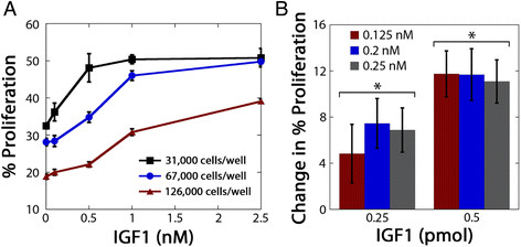
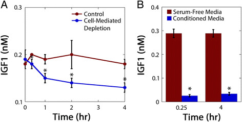
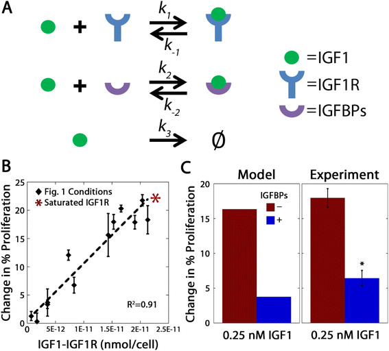
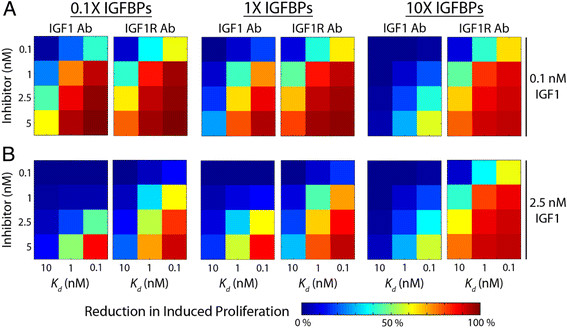
Results: OVCAR5, an immortalized ovarian cancer cell line, were found to be sensitive to IGF1, with the dose of IGF1 (i.e., the total mass of IGF1 available) a more reliable predictor of cell response than ligand concentration. The applied dose of IGF1 was depleted by both cell-secreted IGFBPs and endocytic trafficking, with IGFBPs sequestering up to 90% of the available ligand. To explore how different variables (i.e., IGF1, IGFBPs, and IGF1R levels) impacted cell response, a mass-action steady-state model was developed. Examination of the model revealed that the level of IGF1-IGF1R complexes per cell was directly proportional to the extent of proliferation induced by IGF1. Model analysis suggested, and experimental results confirmed, that IGFBPs present during IGF1 treatment significantly decreased IGF1-mediated proliferation. We utilized this model to assess the efficacy of IGF1 and IGF1R antibodies against different network compositions and determined that IGF1R antibodies were more globally effective due to the receptor-limited state of the network.

Conclusions: Changes that affect IGF1R occupancy have predictable effects on IGF1-induced proliferation and our model captured these effects. Analysis of this model suggests that IGF1R antibodies will be more effective than IGF1 antibodies, although the difference was minimal in conditions with low levels of IGF1 and IGFBPs. Examining how different components of the IGF system influence cell response will be critical to improve our understanding of the IGF signaling network in ovarian cancer.


Figures

Figure 1
Figure 1
OVCAR5 proliferation was dependent on both cell density and IGF1 dose.A, OVCAR5 exhibited concentration-dependent proliferation in response to IGF1 treatment at all three cell densities (31,000, 67,000, 126,000 cells/well); however, the extent of proliferation induced by a set concentration of IGF1 treatment was different at the three cell densities. B, Treatment dose (i.e., pmol of IGF1) impacted the extent of OVCAR5 proliferation while concentration had minimal effect. OVCAR5 were plated at a fixed density (116,000 cells/well) to control for cell confluency, and treatment volumes were varied to result in two doses of IGF1 at three different concentrations. *indicates significantly different (p < 0.05) between doses for each concentration, n = 3 per treatment.
Figure 2
Figure 2
IGF1 availability was regulated by cell-mediated ligand depletion and IGFBP sequestration.A, IGF1 was depleted by OVCAR5 in the absence of IGFBPs. B, The majority of IGF1 added to conditioned media was sequestered by cell-secreted IGFBPs. *indicates significant difference (p < 0.05) from cell-free control for A or from serum-free media control for B, n = 3 per treatment.
Figure 3
Figure 3
IGF1-induced proliferation was a function of steady-state levels of IGF1-IGF1R complexes.A, Diagram of interactions included in the model. B, The computationally-determined concentration of steady-state levels of IGF1-IGF1R complexes exhibited a linear relationship with the experimentally-observed increase in proliferation between IGF1-treated OVCAR5 and vehicle controls. Theoretical saturation of IGF1R is represented by an *. C, Model predictions and experimental results of the effect of IGFBPs on OVCAR5 proliferation in response to IGF1 treatment. The steady-state model predicted that the presence of IGFBPs in the cell culture media would reduce steady-state levels of IGF1-IGF1R complexes and result in decreased cell proliferation. Experimental tests confirmed both the qualitative and quantitative extent of this IGFBP effect. *indicates significant difference (p < 0.05) from IGFBP-negative condition, n = 3 per treatment.
Figure 4
Figure 4
Model-predicted reduction in cell proliferation in response to antibody treatment indicated that IGF1R-blocking antibodies will be more effective than IGF1-neutralizing antibodies. A range of antibody dissociation constants (Kd, 0.1-10 nM) were used to simulate the effect of high to low binding affinity. The effects of the antibody in the presence of three different IGFBP concentrations at A, low (0.1 nM) or B, high (2.5 nM) IGF1 level were determined using the steady-state model. Model results indicated that an antibody that blocks IGF1R would more strongly decrease the steady-state concentration of IGF1-IGF1R complexes and consequently, inhibit IGF1-induced cell proliferation, than an antibody that binds and neutralizes IGF1.

Similar articles

Cited by

References

    1. Le Roith D. Seminars in medicine of the Beth Israel Deaconess Medical Center. Insulin-like growth factors. N Engl J Med. 1997;336:633–640. - PubMed
    1. Weroha SJ, Haluska P. The insulin-like growth factor system in cancer. Endocrinol Metab Clin North Am. 2012;41:335–350. vi. - PMC - PubMed
    1. Baserga R, Peruzzi F, Reiss K. The IGF-1 receptor in cancer biology. Int J Cancer. 2003;107:873–877. - PubMed
    1. Sell C, Dumenil G, Deveaud C, Miura M, Coppola D, DeAngelis T, Rubin R, Efstratiadis A, Baserga R. Effect of a null mutation of the insulin-like growth factor I receptor gene on growth and transformation of mouse embryo fibroblasts. Mol Cell Biol. 1994;14:3604–3612. - PMC - PubMed
    1. Resnicoff M, Abraham D, Yutanawiboonchai W, Rotman HL, Kajstura J, Rubin R, Zoltick P, Baserga R. The insulin-like growth factor I receptor protects tumor cells from apoptosis in vivo. Cancer Res. 1995;55:2463–2469. - PubMed

Publication types