Review
. 2014 Sep 25;124(13):2013-24.
doi: 10.1182/blood-2014-01-453134. Epub 2014 Aug 12.

Src family kinases: at the forefront of platelet activation

Affiliations
Review

Src family kinases: at the forefront of platelet activation

Yotis A Senis et al. Blood. .

Abstract

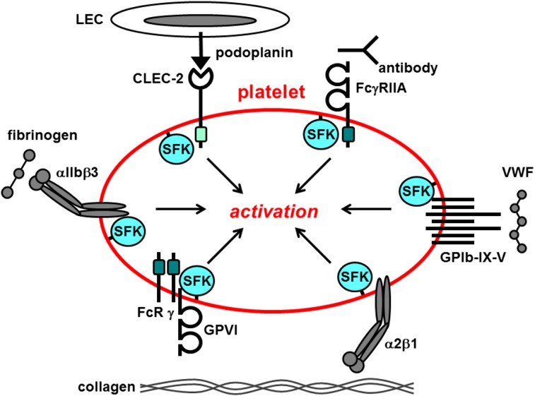
Src family kinases (SFKs) play a central role in mediating the rapid response of platelets to vascular injury. They transmit activation signals from a diverse repertoire of platelet surface receptors, including the integrin αIIbβ3, the immunoreceptor tyrosine-based activation motif-containing collagen receptor complex GPVI-FcR γ-chain, and the von Willebrand factor receptor complex GPIb-IX-V, which are essential for thrombus growth and stability. Ligand-mediated clustering of these receptors triggers an increase in SFK activity and downstream tyrosine phosphorylation of enzymes, adaptors, and cytoskeletal proteins that collectively propagate the signal and coordinate platelet activation. A growing body of evidence has established that SFKs also contribute to Gq- and Gi-coupled receptor signaling that synergizes with primary activation signals to maximally activate platelets and render them prothrombotic. Interestingly, SFKs concomitantly activate inhibitory pathways that limit platelet activation and thrombus size. In this review, we discuss past discoveries that laid the foundation for this fundamental area of platelet signal transduction, recent progress in our understanding of the distinct and overlapping functions of SFKs in platelets, and new avenues of research into mechanisms of SFK regulation. We also highlight the thrombotic and hemostatic consequences of targeting platelet SFKs.


Figures

Figure 1
Figure 1
Src family kinases initiate primary activation in platelets. Src family kinases (SFKs) phosphorylate adaptors, enzymes, and cytoskeletal proteins downstream of a variety of platelet surface receptors that collectively coordinate platelet activation. Dark green box, ITAM; light green box, hemi-ITAM. LEC, lymphatic endothelial cell.
Figure 2
Figure 2
General structure of SFKs. All SFKs share a common structure consisting of an N-terminal myristoyl group attached to an SH4 domain, a unique region, an SH3 domain, an SH2 domain, an SH2-kinase proline-rich linker region, and an SH1 or kinase domain. Yes, Fyn, Fgr, Lyn, Hck, and Lck contain a cysteine residue within the myristoylation peptide sequence that gets palmitoylated and mediates localization to lipid rafts. There is a conserved tyrosine residue in the activation loop and one in the C-terminal tail. Phosphorylation of the activation loop tyrosine by trans-autophosphorylation increases SFK activity, whereas phosphorylation of the C-terminal tyrosine by C-terminal Src kinase (Csk) or the structurally related Csk homologous kinase (Chk) inhibits SFK activity. Dephosphorylation of the activation loop and C-terminal inhibitory tyrosine residues by PTPs attenuates and increases SFK activity, respectively. Green denotes activation, and red denotes inhibition of SFK activity.
Figure 3
Figure 3
SFKs in integrin αIIbβ3 proximal signaling. (A) The integrin αIIbβ3 is in a low-affinity conformation on the surface of resting platelets. The SFKs Src and Fyn constitutively associate with the cytoplasmic tail of the β3 subunit via their SH3 domains. Src is maintained in an inactive conformation by Csk, which forms a complex with β3 and Src. Inside-out signaling induces the integrin to adopt a high-affinity conformation and fibrinogen binding. Csk subsequently dissociates from the complex and is replaced by the nontransmembrane PTP-1B that dephosphorylates the C-terminal inhibitory tyrosine residue of Src and activates it. The receptor-like PTP CD148 plays a major role in maintaining a pool of activated SFKs at the plasma membrane that contribute to αIIbβ3 signaling. Fibrinogen-mediated clustering of the integrin induces trans-autophosphorylation of the activation loop tyrosine residue of SFKs and maximal activation. (B) The ITIM/immunoreceptor tyrosine-based switch motif (ITSM)–containing inhibitory receptors PECAM-1 and G6b-B are phosphorylated by SFKs downstream of αIIbβ3. The SH2 domain–containing nontransmembrane PTPs Shp1 and Shp2 bind to the tandem phosphorylated ITIM/ITSM. The exact contributions of PECAM-1 and G6b-B to αIIbβ3 signaling remain ambiguous and are denoted by the dashed arrow. SFKs also phosphorylate and activate SH2 domain-containing SHIP-1, which forms a complex with SFKs and the β3 tail. SHIP-1 attenuates integrin signaling by dephosphorylating PI3,4,5P3 to PI3,4P2 and disrupting membrane localization of (PLCγ2), which binds to PI3,4,5P3 via its pleckstrin homology (PH) domain. PLCγ2 must associate with the plasma membrane in order to hydrolyze PI4,5P2 to the second messenger’s DAG and IP3 that activate serine/threonine PKC and facilitate Ca2+ mobilization, respectively.
Figure 4
Figure 4
SFKs in GPVI-FcR γ-chain proximal signaling. (A) The SFKs Lyn and Fyn constitutively associate with the proline-rich juxtamembrane region of GPVI via their SH3 domains. This interaction unclamps and activates the SFKs. The receptor-like PTP CD148 maintains the SFKs in an activated state by dephosphorylating their C-terminal inhibitory tyrosine residues. Collagen-mediated clustering of the receptor induces trans-autophosphorylation of the activation loop tyrosine residue and maximal SFK activation. SFKs phosphorylate tandem tyrosine residues in the ITAM-containing FcR γ-chain, which provides a high-affinity docking site for the tandem SH2 domain–containing protein-tyrosine kinase Syk. SFKs also phosphorylate and active Syk. SFKs and Syk phosphorylate downstream effectors and propagate the signal. (B) The ITIM/ITSM-containing inhibitory receptors PECAM-1 and G6b-B inhibit GPVI-FcR γ-chain signaling. Lyn phosphorylates tandem tyrosine residues in the ITIM/ITSM in the cytoplasmic tails of PECAM-1 and G6b-B that provides a high-affinity binding site for the SH2 domain–containing nontransmembrane PTPs Shp1 and Shp2. Lyn associated with the cytoplasmic tail of PECAM-1 facilitates phosphorylation. Lyn also phosphorylates and activates SH2 domain–containing SHIP-1 and PKCδ that form a complex and inhibit dense granule secretion.
Figure 5
Figure 5
SFKs in GPIb-IX-V proximal signaling. The SFKs Src and Lyn associate with the GPIbα subunit and initiate inside-out signaling. Binding of VWF to the extracellular region of GPIbα induces SFK activation and phosphorylation of downstream substrates, including the ITAM-containing FcR γ-chain (FcR γ) and FcγRIIA, both of which act as high-affinity docking sites for the tandem SH2 domain–containing protein-tyrosine kinase Syk. The FcR γ-chain is reported to associate with the GPIbα subunit. However, GPIb-IX-V can signal in an ITAM-independent manner. Fyn and PKCδ associate and reciprocally activate one another, propagating the signal.
Figure 6
Figure 6
Src family kinases in Gq- and Gi-coupled receptor signaling. (Ai) The Gq-coupled PAR-4 signals via the activation of phospholipase β (PLCβ), which hydrolyses PI4,5P2 to the second messengers DAG and IP3, which in turn activate serine/threonine PKC and facilitate Ca2+ mobilization, respectively. The SFK Fyn associates with PKCδ downstream of PLCβ, and they reciprocally activate one another. Increased Ca2+ concentration contributes to SFK activation. SFKs lie upstream of the PI3K/Akt pathway. (Aii) PAR-4 coupled to G12/13 mediates platelet shape change via the RhoA/ROCK/MLC pathway. Fyn activated downstream of G12/13 inhibits signaling via Gq-coupled PAR-4. (B) The Gi-coupled ADP receptor P2Y12 signals by inhibiting adenylate cyclase/cAMP/protein kinase A (PKA) and Gβγ-mediated activation of PI3K/Akt. The SFKs Lyn and Fyn bind directly to the Gα subunit and play a critical role in initiating signaling via the PI3K/Akt pathway.
Figure 7
Figure 7
Tyrosine phosphorylation–mediated regulation of SFK activity. SFK activity is regulated through the phosphorylation of conserved tyrosine residues in the C-terminal tail and activation loop. Phosphorylation of the C-terminal tyrosine residue inhibits SFK activity by mediating formation of an intramolecular interaction with the SH2 domain that blocks the active site. A second intramolecular interaction between the SH3 domain and the proline-rich SH2-kinase linker region maintains the SFK in an inactive conformation. Dephosphorylation of the C-terminal tyrosine residue by the protein-tyrosine phosphatases CD148, PTP-1B, and possibly Shp1, releases the intramolecular interactions and actives the SFK. Maximal activation is achieved through trans-autophosphorylation of the activation loop tyrosine residue. Phosphorylation of the C-terminal inhibitory tyrosine residue by Csk or Csk homologous kinase (Chk) re-establishes the SH2 C-terminal inhibitory phosphotyrosine interaction and returns the SFK to an inactive conformation. Dephosphorylation of the activation loop tyrosine returns the SFK to a basally active state. CD148 may be responsible for dephosphorylating this site in platelets.
Figure 8
Figure 8
Alternative mechanisms of regulation. (A) Established and novel mechanisms of regulating SFK activity in platelets. (B) Unclamping and activation of SFKs by disruption of the intramolecular interactions by a proline-rich (PxxP)/phosphotyrosine-containing protein. Disruption of either the SH3-proline-rich linker or the SH2 C-terminal phosphotyrosine intramolecular interactions facilitates SFK activation, irrespective of whether the C-terminal inhibitory tyrosine residue is phosphorylated. Ser/Thr PPs, protein-phosphatases.

References

    1. Ozaki Y, Asazuma N, Suzuki-Inoue K, Berndt MC. Platelet GPIb-IX-V-dependent signaling. J Thromb Haemost. 2005;3(8):1745–1751. - PubMed
    1. Watson SP, Auger JM, McCarty OJ, Pearce AC. GPVI and integrin alphaIIb β3 signaling in platelets. J Thromb Haemost. 2005;3(8):1752–1762. - PubMed
    1. Nuyttens BP, Thijs T, Deckmyn H, Broos K. Platelet adhesion to collagen. Thromb Res. 2011;127(Suppl 2):S26–S29. - PubMed
    1. Navarro-Núñez L, Langan SA, Nash GB, Watson SP. The physiological and pathophysiological roles of platelet CLEC-2. Thromb Haemost. 2013;109(6):991–998. - PMC - PubMed
    1. Kasirer-Friede A, Kahn ML, Shattil SJ. Platelet integrins and immunoreceptors. Immunol Rev. 2007;218:247–264. - PubMed

Publication types

MeSH terms

Substances