Review
. 2014 Nov;1843(11):2784-2795.
doi: 10.1016/j.bbamcr.2014.08.003. Epub 2014 Aug 9.

Macromolecular transport between the nucleus and the cytoplasm: Advances in mechanism and emerging links to disease

Affiliations
Review

Macromolecular transport between the nucleus and the cytoplasm: Advances in mechanism and emerging links to disease

Elizabeth J Tran et al. Biochim Biophys Acta. 2014 Nov.

Abstract

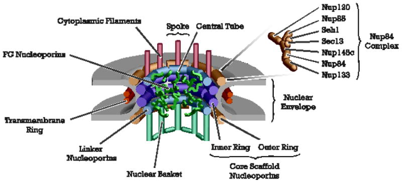
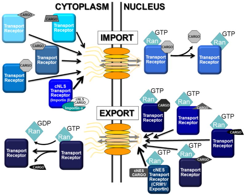
Transport of macromolecules between the cytoplasm and the nucleus is critical for the function of all eukaryotic cells. Large macromolecular channels termed nuclear pore complexes that span the nuclear envelope mediate the bidirectional transport of cargoes between the nucleus and cytoplasm. However, the influence of macromolecular trafficking extends past the nuclear pore complex to transcription and RNA processing within the nucleus and signaling pathways that reach into the cytoplasm and beyond. At the Mechanisms of Nuclear Transport biennial meeting held from October 18 to 23, 2013 in Woods Hole, MA, researchers in the field met to report on their recent findings. The work presented highlighted significant advances in understanding nucleocytoplasmic trafficking including how transport receptors and cargoes pass through the nuclear pore complex, the many signaling pathways that impinge on transport pathways, interplay between the nuclear envelope, nuclear pore complexes, and transport pathways, and numerous links between transport pathways and human disease. The goal of this review is to highlight newly emerging themes in nuclear transport and underscore the major questions that are likely to be the focus of future research in the field.

Keywords: Karyopherin/importin/exportin; Nuclear pore; Nucleocytoplasmic transport; Protein import; RNA export; RNA processing.


Figures

Figure 1
Figure 1. The architecture of the eight-fold, rotationally symmetric nuclear pore complex (provided by Michael Rout)
A schematic diagram of the primary features of the nuclear complex illustrates the sophisticated nature of this macromolecular assembly. The NPC is embedded within a NE pore, formed by fusion of the outer and inner nuclear membranes. A transmembrane ring is present on the outside of the pore, aiding stabilization. Within the core of the NPC are scaffolding proteins that form the inner and outer ring within the channel. At the cytoplasmic and nuclear face are the cytoplasmic filaments and nuclear basket structures, respectively, which serve as docking sites for nuclear transport events. The FG domains of the FG nucleoporins are shown protruding into the aqueous channel of the NPC. These domains serve as docking sites for transport receptors, allowing passage through the pore. The Nup84 complex is a substructure located at the outer periphery of the cytoplasmic face of the NPC, which is required for proper distribution of individual NPCs around the NE [7].
Figure 2
Figure 2. Field emission microscopic images of plant and fungal NPCs (provided by Martin Goldberg)
A. SEM image of the surface of an isolated S. cerevisiae nucleus showing two nuclear pore complexes (green). Arrowheads indicate prominent cytoplasmic filaments. B. SEM image of the surface of a tobacco BY-2 cell nucleus. The channel of the NPCs is indicated by asterisks and the cytoplasmic ring is indicated for one NPC with an arrow. These images illustrate the conservation of the symmetric nature of NPCs and the presence of a cytoplasmic ring and/or filaments across eukaryotes.
Figure 3
Figure 3. Many Pathways Exist for Transport into and out of the Nucleus
The vast majority of nuclear transport depends on the small GTPase Ran and nuclear transport receptors most often termed karyopherins but also importins/exportins. During cargo import (top), transport receptors (shown in shades of blue) associate with cargo (shown in gray) in the cytoplasm through either direct recognition of nuclear localization signals (NLS) or in the case of the classical NLS (cNLS) through the aid of the adaptor protein, importin/karyopherin α (Kap60 in budding yeast). These complexes transit the pore into the nucleus whereby cargo release is triggers by the binding of RanGTP to the receptor-cargo complex. Cargo export (bottom) occurs through formation of an obligate trimeric complex consisting of RanGTP, export cargo (shown in gray/black), and transport receptor (shown in shades of blue). This trimeric complex moves through the pore and is then disassembled in the cytoplasm when Ran hydrolyzes GTP to GDP. The GTPase activity of Ran is specifically localized to the cytoplasm by the presence of GTPase activating proteins. RanGDP is re-imported into the nucleus and then “re-loaded” with GTP through the activity of the Ran guanine nucleotide exchange factor or RanGEF, which is associated with chromatin. The compartmentalization of RanGTP/GDP confers directionality on the system. Thus, there are numerous transport pathways to move macromolecular cargoes into and out of the nucleus. The vast majority of the targeting signals required for these transport pathways have not yet been defined.
Figure 4
Figure 4. Transport selectivity is maintained by the FG barrier within the NPC (adapted from material provided by Roderick Lim and Larisa E. Kapinos)
The question of how the NPC serves as both an efficient barrier and rapid transit machine remains a point of debate. Several models were discussed at the meeting. Prevailing transport models advocate that the barrier mechanism is composed of FG domains. In all cases, selective transport is exclusive to karyopherins (transport receptors; dark green) that bind the FG repeats via multivalent interactions. Small molecules (small red watermarked) diffuse freely through the barrier whereas large non-specific molecules (large red) are withheld due to insufficient interaction with the FG repeats. The models differ with respect to the nature of the FG barrier and its interactions with transport receptors. A. The hydrogel model is based on cohesive interactions between FG repeat domains that create a sieve-like barrier that is selectively permeable to transport receptors [39]. B. The brush model is based on the increased extensibility of the FG Nups due to steric repulsion that wins against competing cohesive interactions [143]. C. The forest model combines aspects of the hydrogel and brush models and is based on the concept that individual FG Nups have distinct properties, being either trees (favoring an extended conformation) or shrubs (favoring a compact conformation). Cohesive interactions also play a role in this model, which suggests that distinct “zones” through the NPC may be taken by individual cargo-transport receptor complexes [144]. D. A Kap-centric NPC barrier mechanism model is based on the ability for the FG domains to bind and accommodate large numbers of Kaps (transport receptors) at physiological concentrations [48, 49]. Owing to strong binding avidity, the Kaps that reside within the FG domains (dark green) are slow and form integral barrier constituents. Nevertheless, this is a prerequisite for weakly-bound Kaps (light green) that dominate fast transport due to limited penetration into the pre-occupied FG domains (e.g., aka the “dirty velcro” effect).
Figure 5
Figure 5. Messenger RNA (mRNA) assembly is required for transport through the NPC (provided by Benoit Palancade)
Export-competent mRNAs are assembled in the nucleus during transcription and mRNA processing steps. This includes binding of transport adaptor proteins such as Yra1, Npl3 and Nab2 to mRNA. These adaptor proteins (shown in gray) recruit the mRNA export receptor heterodimer, Mex67/Mtr2 (TAP/p15 in mammals), and facilitate docking of the mRNA-protein complex (mRNP) with the proteins at the nuclear face of the NPC (i.e., Mlp1/2) [145]. Direct binding of Mex67/Mtr2 to FG repeats within the NPC facilitates movement of the mRNP to the cytoplasm where the activity of the RNA helicase Dbp5 promotes mRNP remodeling and release into the cytoplasm. Proteins that remain associated with the mRNP in the cytoplasm can then influence mRNA stability, cellular localization and/or translational efficiency of the message.
Figure 6
Figure 6. Improved methods for imaging single mRNA molecules in S. cerevisiae (provided by David Grünwald)
A. Budding yeast cells imaged in buffer show high contrast in Phase and DIC. B. Refractive Index (R.I.) differences between immersion and buffer are high for use of high N.A. objectives leading to a loss of emission light. The R.I. of yeast cells is unknown. C. Consequently, at viable excitation light power mRNA fluorescent signal from mRNA in the budding yeast cell is very dim. D & E. Modification of imaging conditions leads to reduced contrast in Phase (DIC contrast does not yield image) resulting in (F) improved mRNA signal for low light imaging.

Similar articles

Cited by

References

    1. Raices M, D'Angelo MA. Nuclear pore complex composition: a new regulator of tissue-specific and developmental functions. Nature reviews Molecular cell biology. 2012;13:687–699. - PubMed
    1. Grossman E, Medalia O, Zwerger M. Functional architecture of the nuclear pore complex. Annual review of biophysics. 2012;41:557–584. - PubMed
    1. Wente SR, Rout MP. The nuclear pore complex and nuclear transport. Cold Spring Harbor perspectives in biology. 2010;2:a000562. - PMC - PubMed
    1. Fernandez-Martinez J, Rout MP. A jumbo problem: mapping the structure and functions of the nuclear pore complex. Current opinion in cell biology. 2012;24:92–99. - PMC - PubMed
    1. Rout MP, Aitchison JD, Suprapto A, Hjertaas K, Zhao Y, Chait BT. The yeast nuclear pore complex: composition, architecture, and transport mechanism. The Journal of cell biology. 2000;148:635–651. - PMC - PubMed

LinkOut - more resources