. 2014 Aug 13:4:6044.
doi: 10.1038/srep06044.

Functional tooth restoration by next-generation bio-hybrid implant as a bio-hybrid artificial organ replacement therapy

Affiliations

Functional tooth restoration by next-generation bio-hybrid implant as a bio-hybrid artificial organ replacement therapy

Masamitsu Oshima et al. Sci Rep. .

Abstract

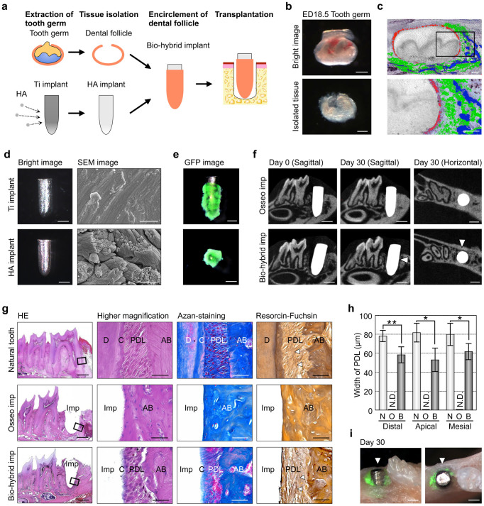
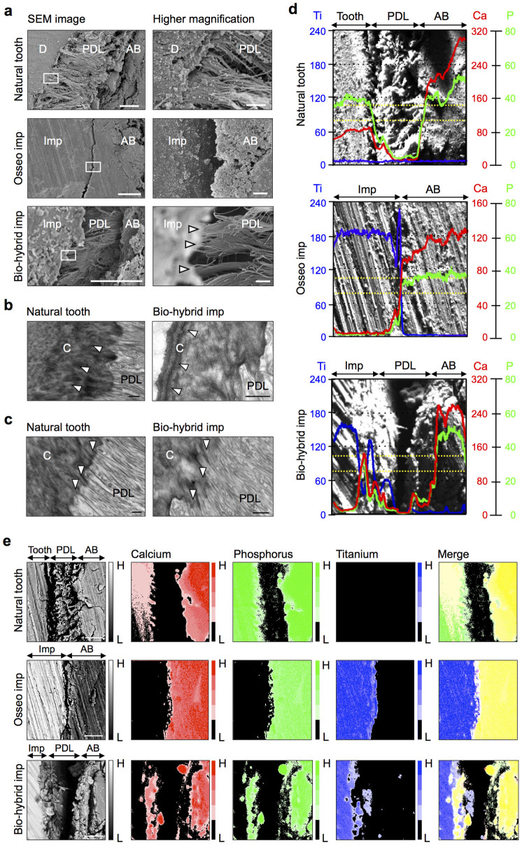
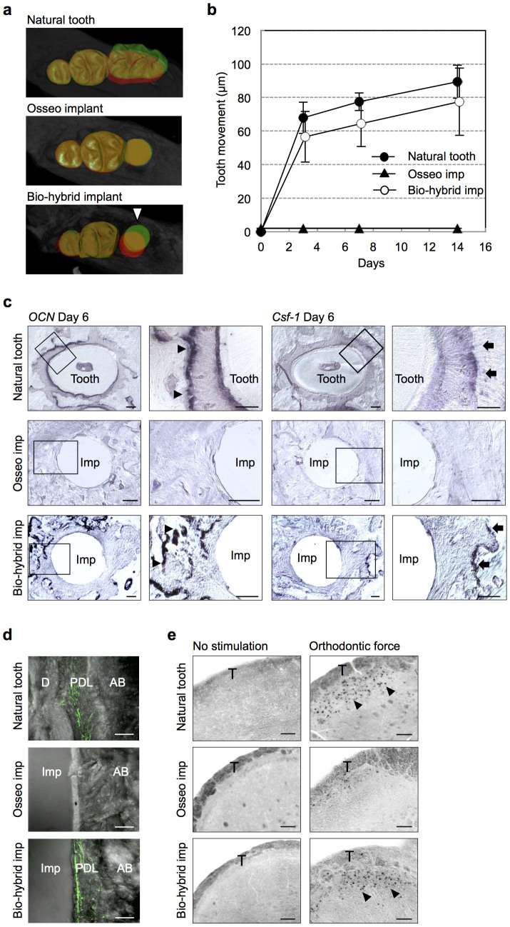
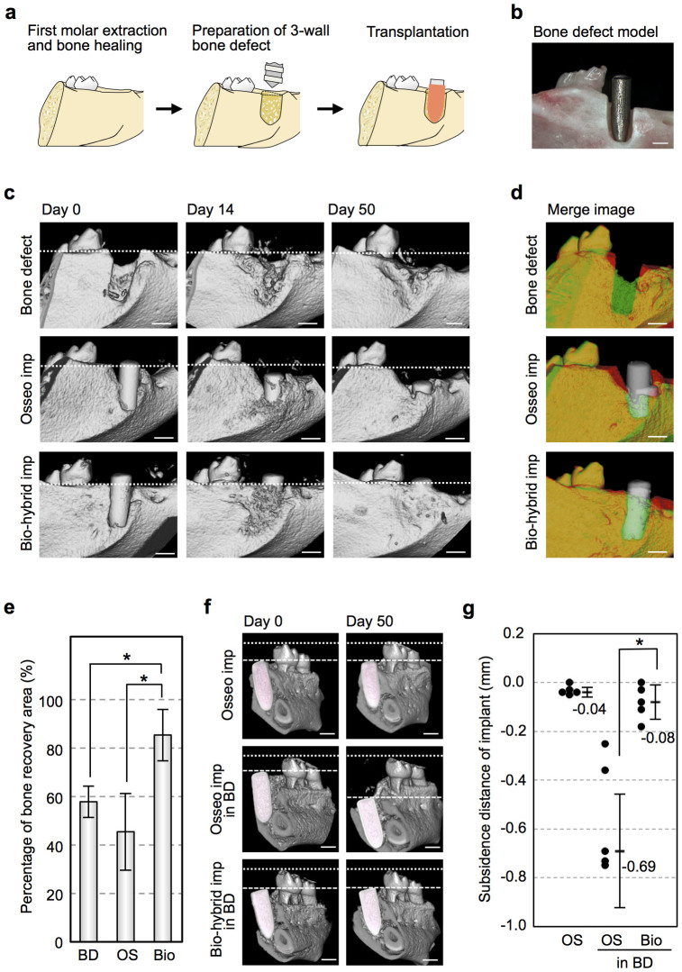
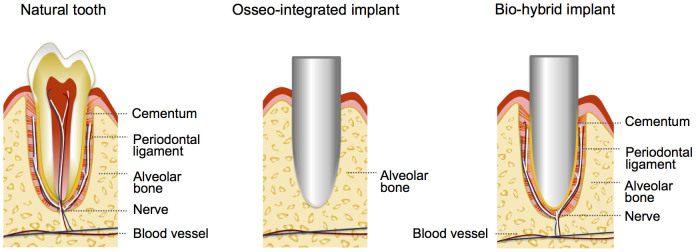
Bio-hybrid artificial organs are an attractive concept to restore organ function through precise biological cooperation with surrounding tissues in vivo. However, in bio-hybrid artificial organs, an artificial organ with fibrous connective tissues, including muscles, tendons and ligaments, has not been developed. Here, we have enveloped with embryonic dental follicle tissue around a HA-coated dental implant, and transplanted into the lower first molar region of a murine tooth-loss model. We successfully developed a novel fibrous connected tooth implant using a HA-coated dental implant and dental follicle stem cells as a bio-hybrid organ. This bio-hybrid implant restored physiological functions, including bone remodelling, regeneration of severe bone-defect and responsiveness to noxious stimuli, through regeneration with periodontal tissues, such as periodontal ligament and cementum. Thus, this study represents the potential for a next-generation bio-hybrid implant for tooth loss as a future bio-hybrid artificial organ replacement therapy.


Figures

Figure 1
Figure 1. Engraftment of a bio-hybrid implant in a tooth loss model.
(a) Schematic representation of the generative technology of bio-hybrid implant. (Drawings by C.T.). (b) Photographs of ED18.5 tooth germ (upper) and isolated dental follicle tissue (lower). Scale bar, 100 μm. (c) Layer arrangement indicated by in situ hybridisation analysis for the expression patterns of F-spondin (red), Periostin (green) and Osteocalcin (blue) in ED18.5 dental follicle tissue. Scale bar, 100 μm. (d) Photographs (left) and surface analysis (right) of titanium implant and HA implant using SEM. Scale bar, 500 μm and 1.0 μm in the photographs and SEM images, respectively. (e) Merged images of a bio-hybrid dental implant using ED18.5 dental follicles isolated from GFP transgenic mice (upper, sagittal view; lower, horizontal view). Scale bar, 500 μm. (f) Micro-CT images of an osseo-integrated implant and a bio-hybrid implant to in sagittal section (left, centre) and horizontal section (right) at transplantation period of Day 0 and Day 30. Bio-hybrid implant images were observed in the periodontal ligament space (arrowhead). Scale bar, 500 μm. (g) Histological analysis of a natural tooth (upper), an engrafted osseo-integrated implant (middle) and an engrafted bio-hybrid implant (lower) at 30 days post-transplantation was performed. HE, Azan, and Resorcin-Fuschin staining are shown. Scale bar, 500 μm in the lower magnification (left column) and 50 μm in the higher magnification (centre-left, centre-right and right column). D, dentin; C, cementum; AB, alveolar bone; PDL, periodontal ligament; Imp, implant. (h) Measurement of the width of periodontal ligament area. The periodontal ligament was not detected in osseo-integrated implants at 30 days post-transplantation. N, natural tooth; O, osseo-integrated implant; B, bio-hybrid implant; N.D., Not detected. Error bars represent the standard deviation (n = 5). *p < 0.05, **p < 0.01 (Mann-Whitney U-test). (i) Photograph of a bio-hybrid implant using ED18.5 dental follicles isolated from GFP transgenic mice at 30 days post-transplantation. Arrowhead, bio-hybrid implant. Scale bar, 500 μm.
Figure 2
Figure 2. Structural analyses of a periodontal tissue in the bio-hybrid implant.
(a) Scanning electron microscopic (SEM) images of natural tooth (upper), the engrafted osseo-integrated implant (middle) and the engrafted bio-hybrid implant (lower) at 30 days post transplantation was performed. Scale bar, 20 μm and 1.0 μm in the lower and higher magnification, respectively. D, dentin; AB, alveolar bone; PDL, periodontal ligament; Imp, implant. (b and c) Transmission electron microscopic (TEM) observation of a natural tooth (left) and the engrafted bio-hybrid implant (right). Formation of lamellar cementum (b, arrowhead) and invasion of Sharpey's fibres into the cementum (c, arrowhead). Scale bar, 500 nm. C, cementum; PDL, periodontal ligament. (d) Amounts of calcium (Ca, red), phosphorus (P, green), and titanium (Ti, blue) in a natural tooth (top), the engrafted osseo-integrated implant (middle) and the engrafted bio-hybrid implant (bottom), as determined by SEM. The amounts of elements were measured in the area between dotted lines. AB, alveolar bone; PDL, periodontal ligament; Imp, implant. (e) Elemental mapping superposition of the natural tooth (top), osseo-integrated implant (middle) and bio-hybrid implant (bottom). Calcium (Ca, red), phosphorus (P, green), titanium (Ti, blue) and merged images are shown. AB, alveolar bone; PDL, periodontal ligament; Imp, implant.
Figure 3
Figure 3. Functional regeneration of a bio-hybrid implant.
(a and b) Horizontal superposition of micro-CT images (left) of the natural tooth (top), osseo-integrated implant (middle) and bio-hybrid implant (bottom) at days 0 (red) and 14 days (green) of experimental orthodontic treatment. The movement distances of tooth and both implants by orthodontic force were measured after experimental orthodontic treatment at days 0, 3, 7 and 14 (right). Data represent the mean ± s.d.; n = 5 for natural tooth, osseo-integrated implant and bio-hybrid implant, respectively. (c) Sections of natural tooth, osseo-integrated and bio-hybrid implants were analysed by in situ hybridization analysis for Ocn and Csf-1 mRNA at day 6 of orthodontic treatment. Ocn mRNA-positive cells (arrowhead) and Csf-1 mRNA-positive cells (arrow) are indicated. Scale bar, 100 μm. (d) Nerve fibres in the PDL of the natural tooth (top), osseo-integrated implant (middle), and bio-hybrid implant (bottom) were analysed by immunohistochemistry using specific antibodies for neurofilament (NF; green). Scale bar, 50 μm. D, dentin; AB, alveolar bone; PDL, periodontal ligament; Imp, implant. (e) Analysis of c-Fos immunoreactive neurons in the medullary dorsal horns of mice after 0 hours (no stimulation, control; left) and 2 hours of stimulation by orthodontic force (right). c-Fos protein (arrowhead) was detectable after stimulation in the natural tooth (top) and bio-hybrid implant (bottom). Scale bar, 100 μm. T, spinal trigeminal tract.
Figure 4
Figure 4. Alveolar bone regeneration by transplantation of a bio-hybrid implant.
(a) Schematic representation of the development of a murine 3-wall bone defect model and transplantation of the implant. (Drawings by C.T.) (b) Photographic representation of a murine 3-wall bone defect model and transplantation of the implant. Scale bar, 500 μm. (c) Micro-CT images of vertical alveolar bone regeneration processes in the bone defect without an implant (top), with transplantation of an osseo-integrated implant (middle) and with the transplantation of a bio-hybrid implant (bottom). Vertical bone formation was observed with the bio-hybrid implant, and the bone had recovered almost completely 50 days after transplantation. The superior edges of the recipient alveolar bone are indicated by a dotted line. Scale bar, 500 μm. (d) Three-dimensional superposition of micro-CT images for the non-treated control (top), the osseo-integrated implant (middle) and the bio-hybrid implant (bottom) in the 3-wall bone defect at 0 (red) and 50 days (green) after transplantation. Scale bar, 500 μm. (e) Regenerated bone area in the buccal region for the non-treated control (bone defect; BD), osseo-integrated implant (OS) and bio-hybrid implant (Bio) after 50 days in the 3-wall bone defect model. The data are presented as the mean ± s.d. with n = 5 for each experimental group. *p < 0.01 (Bonferroni test). (f) Three-dimensional frontal micro-CT images of the subsidence of the osseo-integrated implant into normal tooth loss region (upper), osseo-integrated implant into the bone defect model (middle) and bio-hybrid implant into the bone defect model (lower). The height to the top of the second molar cusp is indicated by the dotted line, and the top of the implant is marked by a dashed line. Scale bar, 500 μm. (g) Vertical subsidence of the osseo-integrated implants in the normal tooth loss region (OS) and in the 3-wall bone defect model (OS in BD) as well as the bio-hybrid implants in the 3-wall bone defect model (Bio in BD) 50 days after transplantation. The data are presented as the median ± s.d. with n = 5 for each experimental group. *p < 0.01 (Mann-Whitney U-test).
Figure 5
Figure 5. Model of the connection to the periodontal tissues of a bio-hybrid implant.
Schematic representation of the natural tooth, osseo-integrated implant and bio-hybrid implant. The bio-hybrid implant restored physiological functions, including bone remodelling, regeneration of severe bone-defect and responsiveness to noxious stimulations, through regeneration with periodontal tissues, such as cementum, PDL and alveolar bone. (Drawings by K.N.).

Similar articles

Cited by

References

    1. Bengel F. M. et al. Effect of sympathetic reinnervation on cardiac performance after heart transplantation. N Engl J Med. 345, 731–738 (2001). - PubMed
    1. Copeland J. G. et al. Cardiac Replacement with a Total Artificial Heart as a Bridge to Transplantation. N Engl J Med. 351, 859–867 (2004). - PubMed
    1. Strain A. J. & Neuberger J. M. A bioartificial liver-state of the art. Science 295, 1005–1009 (2002). - PubMed
    1. Davenport A. et al. A wearable haemodialysis device for patients with end-stage renal failure: a pilot study. Lancet 370, 2005–2010 (2005). - PubMed
    1. Kelly S. K. et al. A hermetic wireless subretinal neurostimulator for vision prostheses. IEEE Trans Biomed Eng. 58, 3195–3205 (2011). - PMC - PubMed

Publication types

MeSH terms

LinkOut - more resources