. 2014 Oct;35(10):1163-70.
doi: 10.1002/humu.22631. Epub 2014 Sep 10.

A mutation in the CASQ1 gene causes a vacuolar myopathy with accumulation of sarcoplasmic reticulum protein aggregates

Affiliations

A mutation in the CASQ1 gene causes a vacuolar myopathy with accumulation of sarcoplasmic reticulum protein aggregates

Daniela Rossi et al. Hum Mutat. 2014 Oct.

Abstract

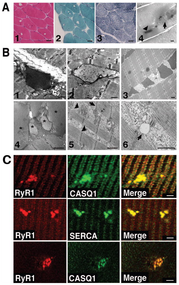
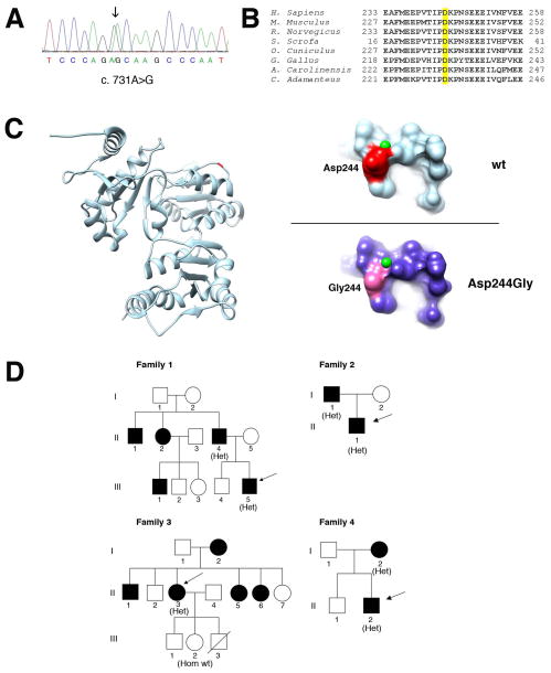
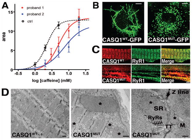
A missense mutation in the calsequestrin-1 gene (CASQ1) was found in a group of patients with a myopathy characterized by weakness, fatigue, and the presence of large vacuoles containing characteristic inclusions resulting from the aggregation of sarcoplasmic reticulum (SR) proteins. The mutation affects a conserved aspartic acid in position 244 (p.Asp244Gly) located in one of the high-affinity Ca(2+) -binding sites of CASQ1 and alters the kinetics of Ca(2+) release in muscle fibers. Expression of the mutated CASQ1 protein in COS-7 cells showed a markedly reduced ability in forming elongated polymers, whereas both in cultured myotubes and in in vivo mouse fibers induced the formation of electron-dense SR vacuoles containing aggregates of the mutant CASQ1 protein that resemble those observed in muscle biopsies of patients. Altogether, these results support the view that a single missense mutation in the CASQ1 gene causes the formation of abnormal SR vacuoles containing aggregates of CASQ1, and other SR proteins, results in altered Ca(2+) release in skeletal muscle fibers, and, hence, is responsible for the clinical phenotype observed in these patients.

Keywords: CASQ1; aggregate myopathy; calsequestrin; sarcoplasmic reticulum; skeletal muscle.


Figures

Figure 1
Figure 1. Morphological characterization of skeletal muscle fibers from patients
A. Hystological analysis. Hematoxilin-eosin (1), Gomori-modified trichrome (2) and NADH-TR (3) stainings of muscle fibers of patient II:1 of family 2. Longitudinal sections from a biopsy of patient II:3 of family 3 stained with Toluidine Blue (4) showing rectangular dark inclusions defined as type 1 (arrowhead), and round pale inclusion defined as type 2 (arrow) (Tomelleri et al.,2006) . Scale bars: panels 1–3 = 40 μm, panel 4 = 5 μm. B. Electron microscope analysis. Representative EM images of type 1 and type 2 inclusions from a biopsy of patient II:1 of family 2 (1 and 2) and of type 1 inclusions from a muscle biopsy of the sporadic patient (3). Representative EM images of patient II:3 of family 3 showing type 2 inclusions labelled with asterisks (4) and areas containing cytoplasmic debris and abnormal mitochondria (arrowheads in 5). The arrow (5) points to a misaligned Z disk. A vacuole clearly connected to the SR membrane of a Ca2+ release units is indicated with an arrow (6). Scale bars = 1 μm. C. Immunofluorescence analysis. Single muscle fibers (upper and middle panels) or cross sections (lower panels) of skeletal muscle biopsy of patient I:1 of family 2 stained with antibodies against CASQ1, SERCA andRyR1. Merged images show co-localization of CASQ1, RyR1 and SERCA in the aggregates. Scale bars = 2 μm.
Figure 2
Figure 2. Genetic and molecular analysis of the p.Asp244Gly mutation
A. Electropherogram of the CASQ1 gene sequence of one patient. The patient is heterozygous for the c.731A>G variant (arrow). B. Phylogenetic alignment of the CASQ1 orthologs. The aspartic acid in position 244 in the human CASQ1 is highlighted yellow and is conserved in species ranging from snakes (C. Adamanteus) to humans. C. 3D structure of human CASQ1 protein. Ribbon representation of the model of chain A of the human CASQ1 3D structure (PDB:3UOM) with the Asp244 shown in red/dark (left). On the right, detailed views of predicted surface structure of the local molecular environments surrounding the Asp244 (upper panel) or the Gly244 residues (lower panel). The small (green) spheres represent Ca2+ ions. D. Pedigrees of families. Black filled symbols represent affected family members. The genotype of individuals is shown in parenthesis as follows: Het, heterozygous for the c.731A>G variant; Hom wt, homozygous for the wild-type allele. Arrows indicate probands.
Figure 3
Figure 3. Ca2+ release kinetics in muscle fibers of two patients and expression of recombinant CASQ1WT and CASQ1MUT proteins
A. Ca2+ release kinetics in muscle fibers of two patients. Dose-response curves of permeabilized muscle fibers from control healthy subjects (black dashed line, average of 48 fibers from 4 subjects), patient I:1 of family 2 (continuous red line, average of 15 fibers) and from the sporadic case (continuous blue line, average of 21 fibers) exposed to increasing concentrations of caffeine (from 0.1 to 20 mM). Curves are interpolated by a sigmoidal Hill equation: Y=B+(Amax-B)/(1+10^((LogEC50-X)n)). The parameter logEC50 is significantly greater (p<0.01) in the patient I:1 of family 2 (0.697+/− 0.062) and in the sporadic case (0.863+/− 0.101) than in controls (0.343 +/− 0.006). B. CASQ1WT-GFP and CASQ1MUT-GFP were expressed in COS-7 cells. Living cells were imaged by confocal laser scan microscopy. Scale bars = 7 μm. C. Rat myotubes expressing recombinant CASQ1WT and CASQ1MUT were immunostained with antibodies against CASQ1 and RyR1. Scale bar = 2 μm. D. EM analysis of CASQ1WT and CASQ1MUT in mouse FDB fibers. Fibers expressing CASQ1MUT show the presence of vacuoles filled with electron-dense material (asterisks). These vacuoles were not present in fibers expressing CASQ1WT. Higher magnification shows enlarged vesicles derived from the SR terminal cisternae: arrows point to RyR-feet between an enlarged SR vesicle and a T-tubule. M, mitochondrion; TT, transverse tubule; SR, sarcoplasmic reticulum. Scale bars in left, middle and right panels = 0.5, 1 and 0.1 μm, respectively.

References

    1. Boncompagni S, d’Amelio L, Fulle S, Fanò G, Protasi F. Progressive disorganization of the excitation-contraction coupling apparatus in ageing human skeletal muscle as revealed by electron microscopy: a possible role in the decline of muscle performance. J Gerontol A Biol Sci Med Sci. 2006;61:995–1008. - PubMed
    1. Cho JH, Ko KM, Singaruvelu G, Lee W, Kang GB, Rho SH, Park BJ, Yu JR, Kagawa H, Eom SH, Kim do H, Ann J. Functional importance of polymerization and localization of calsequestrin in C. elegans. J Cell Sci. 2007;120:1551–1558. - PubMed
    1. Dainese M, Quarta M, Lyfenko AD, Paolini C, Canato M, Reggiani C, Dirksen RT, Protasi F. Anesthetic- and heat-induced sudden death in calsequestrin-1-knockout mice. FASEB J. 2009;23:1710–1720. - PMC - PubMed
    1. Damiani E, Volpe P, Margreth A. Coexpression of two isoforms of calsequestrin in rabbit slow-twitch muscle. J Muscle Res Cell Motil. 1990;11:522–530. - PubMed
    1. Doria C, Toniolo L, Verratti V, Cancellara P, Pietrangelo T, Marconi V, Paoli A, Pogliaghi S, Fano G, Reggiani C, Capelli C. Improved VO2 uptake kinetics and shift in muscle fiber type in high altitude trekkers. J Appl Physiol. 2011;111:1597–605. - PubMed

Publication types

MeSH terms

Supplementary concepts