Review
. 2014 Nov 15;32(7):1256-1268.
doi: 10.1016/j.biotechadv.2014.07.009. Epub 2014 Aug 10.

Three-dimensional in vitro tumor models for cancer research and drug evaluation

Affiliations
Review

Three-dimensional in vitro tumor models for cancer research and drug evaluation

Xian Xu et al. Biotechnol Adv. .

Abstract

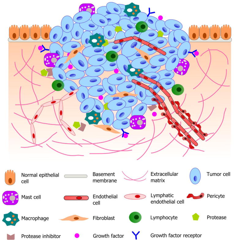
Cancer occurs when cells acquire genomic instability and inflammation, produce abnormal levels of epigenetic factors/proteins and tumor suppressors, reprogram the energy metabolism and evade immune destruction, leading to the disruption of cell cycle/normal growth. An early event in carcinogenesis is loss of polarity and detachment from the natural basement membrane, allowing cells to form distinct three-dimensional (3D) structures that interact with each other and with the surrounding microenvironment. Although valuable information has been accumulated from traditional in vitro studies in which cells are grown on flat and hard plastic surfaces (2D culture), this culture condition does not reflect the essential features of tumor tissues. Further, fundamental understanding of cancer metastasis cannot be obtained readily from 2D studies because they lack the complex and dynamic cell-cell communications and cell-matrix interactions that occur during cancer metastasis. These shortcomings, along with lack of spatial depth and cell connectivity, limit the applicability of 2D cultures to accurate testing of pharmacologically active compounds, free or sequestered in nanoparticles. To recapitulate features of native tumor microenvironments, various biomimetic 3D tumor models have been developed to incorporate cancer and stromal cells, relevant matrix components, and biochemical and biophysical cues, into one spatially and temporally integrated system. In this article, we review recent advances in creating 3D tumor models employing tissue engineering principles. We then evaluate the utilities of these novel models for the testing of anticancer drugs and their delivery systems. We highlight the profound differences in responses from 3D in vitro tumors and conventional monolayer cultures. Overall, strategic integration of biological principles and engineering approaches will both improve understanding of tumor progression and invasion and support discovery of more personalized first line treatments for cancer patients.

Keywords: 3D tumor models; Bioreactors; Cancer therapeutics; Drug delivery; Drug resistance; Hydrogels; Microfluidic devices; Scaffolds.


Figures

Fig. 1
Fig. 1
Schematic illustration of a typical tumor microenvironment. Cancer cells reside in a complex microenvironment containing various supporting cells, extracellular matrix (ECM) and a suite of signaling molecules. These environmental components collectively contribute to the tumor-stromal interaction and tumor progression. Adapted from (Joyce and Pollard, 2009; Koontongkaew, 2013).
Fig. 2
Fig. 2
Representative examples of culture platforms employed for the growth of 3D tumoroids. (A) Rotating wall vessel (RWV) bioreactors (i, ii) have been used for the 3D culture of human breast ductal carcinoma cells (T-47D, iii) and glioblastoma cells (labeled with green fluorescent protein, iv). Cells cultured in a state of free fall with minimal shear (ii) organized into millimeter sized aggregates with multiple layers of cells (iii and iv, scale bar: 200 μm). Reproduced with permission (Becker and Souza, 2013), Copyright 2013, Macmillan Publishers Limited. (B) A microfluidic device (i–iii) has been designed for the 3D culture of PC-3 prostate cancer cells. The device is composed of upper and lower channels separated by a non-adhesive, semi-permeable membrane. PC-3 cells, mixed with pre-osteoblasts and endothelial cells and introduced to the device as a monolayer, self-organized into 3D spheroids in one day (iv: optical image; v: fluorescent image, red-PC-3 cells, green-live cells; scale bar: 200 μm). Reproduced with permission (Hsiao et al., 2009), Copyright 2009, Elsevier Ltd. (C) A bilayer hydrogel platform (i) for the 3D culture of prostate cancer cells. LNCaP cells culture in the bottom layer, receiving heparin-binding epidermal growth factor-like growth factor (HB-EGF) released from the top layer, grew into spheroids (ii, iii, nuclei: blue) displaying cortical F-actin (green) and expressing E-cadherin (red). Reproduced with permission (Xu et al., 2012), Copyright 2012, Elsevier Ltd.
Fig. 3
Fig. 3
Comparison of intracellular localization and cell-killing capacity of free Dox and Dox-NPs in 2D and 3D cultures of LNCaP prostate cancer cells. HA-based hydrogels were used for 3D cultures. (A) Confocal images show differential localizations of Dox and Dox-NPs in LNCaP cells after 2 h of drug exposure. Dox is inherently fluorescent and the internalized Dox or Dox-NPs were detected using a Zeiss510 NLO confocal microscope (excitation wavelength at 488 nm with a band pass filter of 565–615 nm). (B) Dose-dependent cell death induced by Dox or Dox-NPs on 2D and 3D cultures of LNCaP prostate cancer cells. Cell apoptosis analysis was performed using Cell Death Detection ELISAPLUS. (C) Summary of the IC50 values for various drug/culture combinations. (*, significant difference compared to the IC50 value of 2D Dox condition, p < 0.05). Reproduced with permission (Xu et al., 2014), Copyright 2014, Elsevier Ltd.
Fig. 3
Fig. 3
Comparison of intracellular localization and cell-killing capacity of free Dox and Dox-NPs in 2D and 3D cultures of LNCaP prostate cancer cells. HA-based hydrogels were used for 3D cultures. (A) Confocal images show differential localizations of Dox and Dox-NPs in LNCaP cells after 2 h of drug exposure. Dox is inherently fluorescent and the internalized Dox or Dox-NPs were detected using a Zeiss510 NLO confocal microscope (excitation wavelength at 488 nm with a band pass filter of 565–615 nm). (B) Dose-dependent cell death induced by Dox or Dox-NPs on 2D and 3D cultures of LNCaP prostate cancer cells. Cell apoptosis analysis was performed using Cell Death Detection ELISAPLUS. (C) Summary of the IC50 values for various drug/culture combinations. (*, significant difference compared to the IC50 value of 2D Dox condition, p < 0.05). Reproduced with permission (Xu et al., 2014), Copyright 2014, Elsevier Ltd.

Similar articles

Cited by

References

    1. Becker JL, Souza GR. Using space-based investigations to inform cancer research on Earth. Nat Rev Cancer. 2013;13:315–27. - PubMed
    1. Bissell MJ, Hines WC. Why don’t we get more cancer? A proposed role of the microenvironment in restraining cancer progression. Nat Med (N Y, NY, U S) 2011;17:320–9. - PMC - PubMed
    1. Bourguignon LYW, Singleton PA, Zhu HB, Diedrich F. Hyaluronan-mediated CD44 interaction with RhoGEF and Rho kinase promotes Grb2-associated binder-1 phosphorylation and phosphatidylinositol 3-kinase signaling leading to cytokine (Macrophage-Colony stimulating factor) production and breast tumor progression. J Biol Chem. 2003;278:29420–34. - PubMed
    1. Bourguignon LYW, Singleton PA, Zhu HB, Zhou B. Hyaluronan promotes signaling interaction between CD44 and the transforming growth factor beta receptor I in metastatic breast tumor cells. J Biol Chem. 2002;277:39703–12. - PubMed
    1. Buchanan CF, Voigt EE, Szot CS, Freeman JW, Vlachos PP, Rylander MN. Three-dimensional microfluidic collagen hydrogels for investigating flow-mediated tumor-endothelial signaling and vascular organization. Tissue Eng. 2014;20:64–75. - PMC - PubMed

Publication types

Substances