Review
. 2014 Aug;115(2-3):198-212.
doi: 10.1016/j.pbiomolbio.2014.08.005. Epub 2014 Aug 10.

Images as drivers of progress in cardiac computational modelling

Affiliations
Review

Images as drivers of progress in cardiac computational modelling

Pablo Lamata et al. Prog Biophys Mol Biol. 2014 Aug.

Abstract

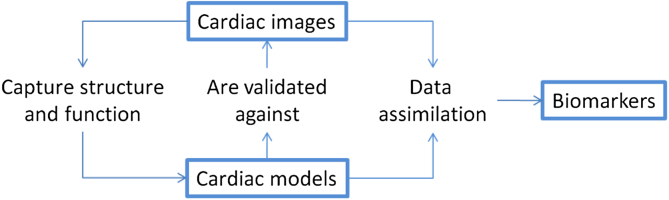
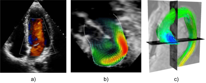
Computational models have become a fundamental tool in cardiac research. Models are evolving to cover multiple scales and physical mechanisms. They are moving towards mechanistic descriptions of personalised structure and function, including effects of natural variability. These developments are underpinned to a large extent by advances in imaging technologies. This article reviews how novel imaging technologies, or the innovative use and extension of established ones, integrate with computational models and drive novel insights into cardiac biophysics. In terms of structural characterization, we discuss how imaging is allowing a wide range of scales to be considered, from cellular levels to whole organs. We analyse how the evolution from structural to functional imaging is opening new avenues for computational models, and in this respect we review methods for measurement of electrical activity, mechanics and flow. Finally, we consider ways in which combined imaging and modelling research is likely to continue advancing cardiac research, and identify some of the main challenges that remain to be solved.

Keywords: Computational cardiac physiology; Medical imaging.


Figures

Fig. 1
Fig. 1
Conceptual scheme of how cardiac images interact with computational models to generate novel insight and drive research progress.
Fig. 2
Fig. 2
Sample slices in three orthogonal directions from an ex vivo 3D MRI acquisition of a rabbit heart, from Bishop et al. (2010b). The original resolution of the images is 26.4 μm × 26.4 μm × 24.4 μm. LV, RV, LA, RA: Left (right) ventricle (atrium). AO: Aorta.
Fig. 3
Fig. 3
Imaging techniques to capture cardiac electrophysiological information. (a) Electro-anatomical map of the atrium, obtained by intra-cardiac catheter approaches, showing a re-entry circuit (Chubb et al., 2014; image courtesy of Dr. H. Chubb, KCL); (b) Functional MRI of the heart capturing the extent of fibrosis for ablation therapy of atrial arrhythmias (top), automatically segmented and reconstructed (bottom; Karim et al., 2014; image courtesy of Dr. R. Karim, KCL). AO: Aorta, LA: Left Atrium, RSV: Right Subclavian Vein, LIV: Left Innominate Vein. (c) Multi-electrode cardiac sock for human epicardiac potential mapping (Nash et al. 2006; image courtesy of Dr. M. Nash, University of Auckland); (d) Optical mapping set-up for ex vivo Langendorff-perfused rabbit heart (Bishop et al. 2014; image courtesy of Dr. R. Burton and Dr. G. Bub, University of Oxford).
Fig. 4
Fig. 4
Illustration of techniques used to capture the mechanical information available in images. (a) Computational mesh of the left ventricle fitted to the domain of the myocardium from the end-diastolic frame of a MRI dynamic short axis stack (Lamata et al., 2011). (b) Tagged MRI frame of a short axis view of the left ventricle with an overlay of a colour encoded deformation field estimated from it by image registration (Chandrashekara et al., 2004). (c) Computational mesh fitted to in vivo DT-MRI data (Toussaint et al., 2013) at two instants of the cardiac cycle, systole (left) and diastole (right), with vectors pointing in the direction of the first eigenvector (fibre), colour encoded with respect to the elevation angle (red to blue, +45 to −45). (d) Echocardiographic speckle tracking (Ledesma-Carbayo et al., 2005). (e) Echocardiography-based electromechanical wave imaging (EWI), illustrating the motion maps (left) and the EWI isochrones (right; both colour coded from 0 ms, red, to 300 ms, blue; Provost et al., 2011b). (f) Elastography by the methods described in (Robert et al., 2009): the panel illustrates the shear modulus in a healthy volunteer at two time points of the cardiac cycle (image courtesy of Dr. R. Sinkus, KCL).
Fig. 5
Fig. 5
Illustration of three main imaging technologies used to capture information on blood flow. (a) 2D Doppler echocardiography is widely used in the clinic; (b) 3D flow reconstructed in an infant with mitral stenosis using registration of multiple 3D Doppler acquisitions (Gómez et al., 2013) (c) Phase-contrast MRI, also known as 4D flow data, colour coded by the pressure maps computed from blood flow velocity data (Krittian et al., 2012) in a chronic dissection case.

References

    1. Aguado-Sierra J., Krishnamurthy A., Villongco C. Patient-specific modeling of dyssynchronous heart failure: a case study. Prog. Biophys. Mol. Biol. 2011;107:147–155. doi: 10.1016/j.pbiomolbio.2011.06.014. - DOI - PMC - PubMed
    1. Ainslie M., Miller C., Brown B., Schmitt M. Cardiac MRI of patients with implanted electrical cardiac devices. Heart. 2014;100:363–369. doi: 10.1136/heartjnl-2013-304324. - DOI - PubMed
    1. Arevalo H., Plank G., Helm P. Tachycardia in post-infarction hearts: insights from 3D image-based ventricular models. PLoS One. 2013;8:e68872. doi: 10.1371/journal.pone.0068872. - DOI - PMC - PubMed
    1. Ashikaga H., Arevalo H., Vadakkumpadan F. Feasibility of image-based simulation to estimate ablation target in human ventricular arrhythmia. Heart Rhythm. 2013;10:1109–1116. doi: 10.1016/j.hrthm.2013.04.015. - DOI - PMC - PubMed
    1. Aslanidi O.V., Nikolaidou T., Zhao J. Application of micro-computed tomography with iodine staining to cardiac imaging, segmentation, and computational model development. IEEE Trans. Med. Imaging. 2013;32:8–17. doi: 10.1109/TMI.2012.2209183. - DOI - PMC - PubMed

Publication types

MeSH terms