

Interrogation of living myocardium in multiple static deformation states with diffusion tensor and diffusion spectrum imaging
- PMID: 25117498
- PMCID: PMC4210665
- DOI: 10.1016/j.pbiomolbio.2014.08.002
Interrogation of living myocardium in multiple static deformation states with diffusion tensor and diffusion spectrum imaging
Abstract
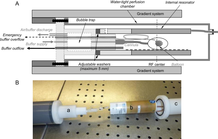
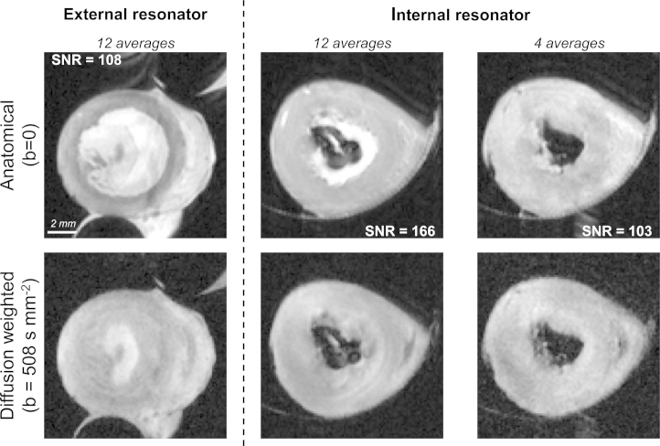
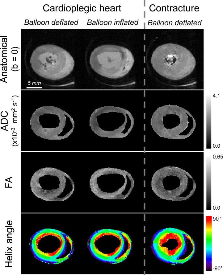
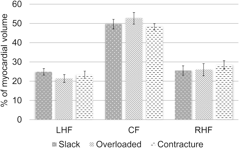
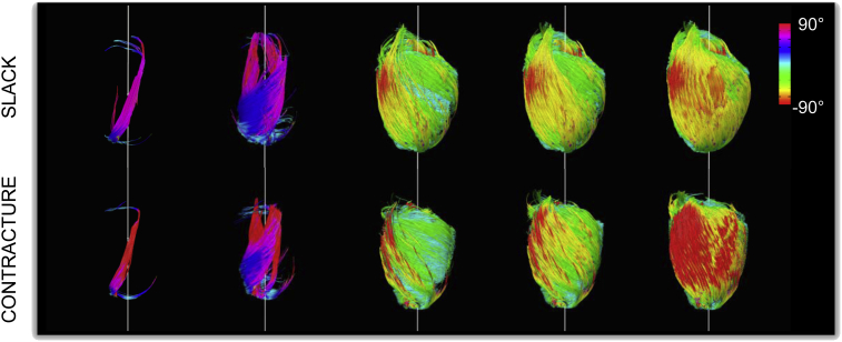
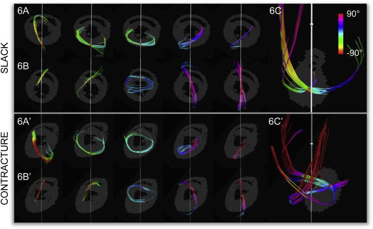
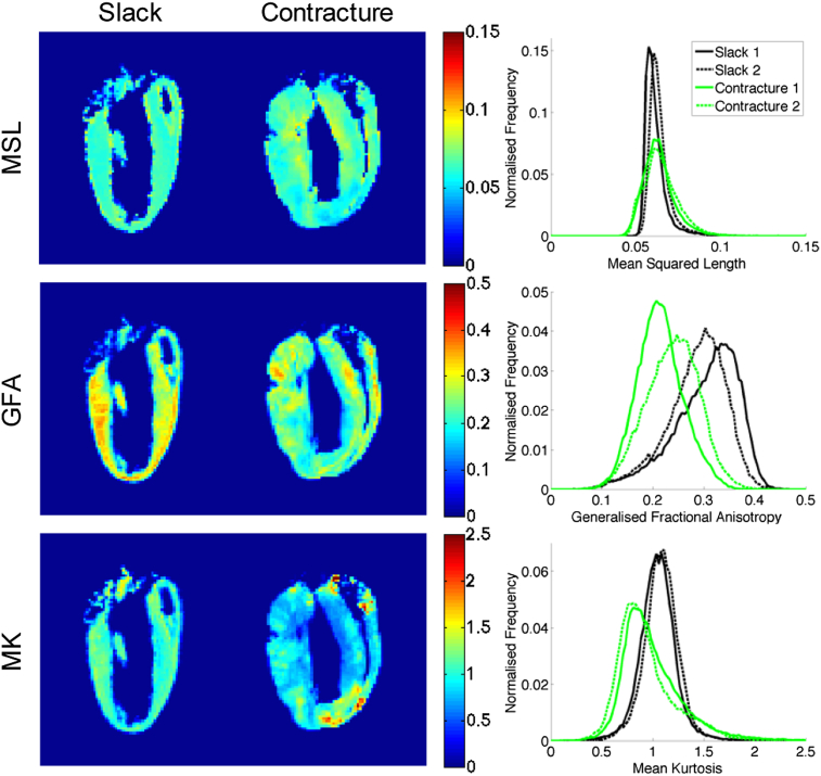
Diffusion tensor magnetic resonance imaging (MRI) reveals valuable insights into tissue histo-anatomy and microstructure, and has steadily gained traction in the cardiac community. Its wider use in small animal cardiac imaging in vivo has been constrained by its extreme sensitivity to motion, exaggerated by the high heart rates usually seen in rodents. Imaging of the isolated heart eliminates respiratory motion and, if conducted on arrested hearts, cardiac pulsation. This serves as an important intermediate step for basic and translational studies. However, investigating the micro-structural basis of cardiac deformation in the same heart requires observations in different deformation states. Here, we illustrate the imaging of isolated rat hearts in three mechanical states mimicking diastole (cardioplegic arrest), left-ventricular (LV) volume overload (cardioplegic arrest plus LV balloon inflation), and peak systole (lithium-induced contracture). An optimised MRI-compatible Langendorff perfusion setup with the radio-frequency (RF) coil integrated into the wet chamber was developed for use in a 9.4T horizontal bore scanner. Signal-to-noise ratio improved significantly, by 75% compared to a previous design with external RF coil, and stability tests showed no significant changes in mean T1, T2 or LV wall thickness over a 170 min period. In contracture, we observed a significant reduction in mean fractional anisotropy from 0.32 ± 0.02 to 0.28 ± 0.02, as well as a significant rightward shift in helix angles with a decrease in the proportion of left-handed fibres, as referring to the locally prevailing cell orientation in the heart, from 24.9% to 23.3%, and an increase in the proportion of right-handed fibres from 25.5% to 28.4%. LV overload, in contrast, gave rise to a decrease in the proportion of left-handed fibres from 24.9% to 21.4% and an increase in the proportion of right-handed fibres from 25.5% to 26.0%. The modified perfusion and coil setup offers better performance and control over cardiac contraction states. We subsequently performed high-resolution diffusion spectrum imaging (DSI) and 3D whole heart fibre tracking in fixed ex vivo rat hearts in slack state and contracture. As a model-free method, DSI augmented the measurements of water diffusion by also informing on multiple intra-voxel diffusion orientations and non-Gaussian diffusion. This enabled us to identify the transition from right- to left-handed fibres from the subendocardium to the subepicardium, as well as voxels in apical regions that were traversed by multiple fibres. We observed that both the mean generalised fractional anisotropy and mean kurtosis were lower in hearts in contracture compared to the slack state, by 23% and 9.3%, respectively. While its heavy acquisition burden currently limits the application of DSI in vivo, ongoing work in acceleration techniques may enable its use in live animals and patients. This would provide access to the as yet unexplored dimension of non-Gaussian diffusion that could serve as a highly sensitive marker of cardiac micro-structural integrity.
Keywords: Cardiac MRI; Diffusion spectrum imaging; Diffusion tensor imaging; Isolated heart; Langendorff perfusion; Magnetic resonance imaging.
Copyright © 2014 The Authors. Published by Elsevier Ltd.. All rights reserved.
Figures
Similar articles
-
Histo-anatomical structure of the living isolated rat heart in two contraction states assessed by diffusion tensor MRI.Prog Biophys Mol Biol. 2012 Oct-Nov;110(2-3):319-30. doi: 10.1016/j.pbiomolbio.2012.07.014. Epub 2012 Aug 7. Prog Biophys Mol Biol. 2012. PMID: 23043978 Free PMC article.
-
Spin echo versus stimulated echo diffusion tensor imaging of the in vivo human heart.Magn Reson Med. 2016 Sep;76(3):862-72. doi: 10.1002/mrm.25998. Epub 2015 Oct 7. Magn Reson Med. 2016. PMID: 26445426 Free PMC article.
-
Optimized radiofrequency coil setup for MR examination of living isolated rat hearts in a horizontal 9.4T magnet.Magn Reson Med. 2015 Jun;73(6):2398-405. doi: 10.1002/mrm.25369. Epub 2014 Jul 12. Magn Reson Med. 2015. PMID: 25045897
-
Cardiac Diffusion: Technique and Practical Applications.J Magn Reson Imaging. 2020 Aug;52(2):348-368. doi: 10.1002/jmri.26912. Epub 2019 Sep 4. J Magn Reson Imaging. 2020. PMID: 31482620 Review.
-
Diffusion MR tractography of the heart.J Cardiovasc Magn Reson. 2009 Nov 13;11(1):47. doi: 10.1186/1532-429X-11-47. J Cardiovasc Magn Reson. 2009. PMID: 19912654 Free PMC article. Review.
Cited by
-
A tomographic microscopy-compatible Langendorff system for the dynamic structural characterization of the cardiac cycle.Front Cardiovasc Med. 2022 Dec 22;9:1023483. doi: 10.3389/fcvm.2022.1023483. eCollection 2022. Front Cardiovasc Med. 2022. PMID: 36620622 Free PMC article.
-
Validation of diffusion tensor MRI measurements of cardiac microstructure with structure tensor synchrotron radiation imaging.J Cardiovasc Magn Reson. 2017 Mar 10;19(1):31. doi: 10.1186/s12968-017-0342-x. J Cardiovasc Magn Reson. 2017. PMID: 28279178 Free PMC article.
-
Diffusion tensor imaging and histology of developing hearts.NMR Biomed. 2016 Oct;29(10):1338-49. doi: 10.1002/nbm.3576. Epub 2016 Aug 3. NMR Biomed. 2016. PMID: 27485033 Free PMC article.
-
Development of a cardiovascular magnetic resonance-compatible large animal isolated heart model for direct comparison of beating and arrested hearts.NMR Biomed. 2022 Jul;35(7):e4692. doi: 10.1002/nbm.4692. Epub 2022 Feb 12. NMR Biomed. 2022. PMID: 35040195 Free PMC article.
-
Finite-Element Extrapolation of Myocardial Structure Alterations Across the Cardiac Cycle in Rats.J Biomech Eng. 2015 Oct;137(10):101010. doi: 10.1115/1.4031419. J Biomech Eng. 2015. PMID: 26299478 Free PMC article.
References
-
- Allis J.L., Snaith C.D., Seymour A.L., Radda G.K. 87Rb NMR studies of the perfused rat heart. FEBS Lett. 1989;242(2):215–217. - PubMed
-
- Assaf Y., Freidlin R.Z., Rohde G.K., Basser P.J. New modeling and experimental framework to characterise hindered and restricted water diffusion in brain white matter. Magnetic Reson. Med. 2004;52(5):965–978. - PubMed
-
- Atkinson D.J., Burstein D., Edelman R.R. First-pass cardiac perfusion: evaluation with ultrafast MR imaging. Radiology. 1990;174(3 Pt I):757–762. - PubMed
-
- Bailey I.A., Gadian D.G., Matthews P.M., Radda G.K., Seeley P.J. Studies of metabolism in the isolated, perfused rat heart using 13C NMR. FEBS Lett. 1981;123(2):315–318. - PubMed
Publication types
MeSH terms
Grants and funding
LinkOut - more resources
Full Text Sources
Other Literature Sources
Research Materials
