. 2014 Aug 12;9(8):e104157.
doi: 10.1371/journal.pone.0104157. eCollection 2014.

Phosphoproteomic profiling of human myocardial tissues distinguishes ischemic from non-ischemic end stage heart failure

Affiliations

Phosphoproteomic profiling of human myocardial tissues distinguishes ischemic from non-ischemic end stage heart failure

Matthew A Schechter et al. PLoS One. .

Abstract

The molecular differences between ischemic (IF) and non-ischemic (NIF) heart failure are poorly defined. A better understanding of the molecular differences between these two heart failure etiologies may lead to the development of more effective heart failure therapeutics. In this study extensive proteomic and phosphoproteomic profiles of myocardial tissue from patients diagnosed with IF or NIF were assembled and compared. Proteins extracted from left ventricular sections were proteolyzed and phosphopeptides were enriched using titanium dioxide resin. Gel- and label-free nanoscale capillary liquid chromatography coupled to high resolution accuracy mass tandem mass spectrometry allowed for the quantification of 4,436 peptides (corresponding to 450 proteins) and 823 phosphopeptides (corresponding to 400 proteins) from the unenriched and phospho-enriched fractions, respectively. Protein abundance did not distinguish NIF from IF. In contrast, 37 peptides (corresponding to 26 proteins) exhibited a ≥ 2-fold alteration in phosphorylation state (p<0.05) when comparing IF and NIF. The degree of protein phosphorylation at these 37 sites was specifically dependent upon the heart failure etiology examined. Proteins exhibiting phosphorylation alterations were grouped into functional categories: transcriptional activation/RNA processing; cytoskeleton structure/function; molecular chaperones; cell adhesion/signaling; apoptosis; and energetic/metabolism. Phosphoproteomic analysis demonstrated profound post-translational differences in proteins that are involved in multiple cellular processes between different heart failure phenotypes. Understanding the roles these phosphorylation alterations play in the development of NIF and IF has the potential to generate etiology-specific heart failure therapeutics, which could be more effective than current therapeutics in addressing the growing concern of heart failure.


Conflict of interest statement

Competing Interests: The authors have declared no competing interests exist.

Figures

Figure 1
Figure 1. Experimental Approach.
A: Illustration of the sample preparation and MS work flow. B: Reproducibility/Internal Control. Relative levels of the ADH1 protein spiked into each heart lysate as determined by chromatographic peak intensity. C: Venn Diagram demonstrating the protein and phosphoprotein yield and overlap.
Figure 2
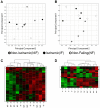
Figure 2. Principal Components Analysis and Hierarchical Clustering.
The expression data for all peptides from the unenriched (A) and phosphopeptide enriched (B) samples were used to perform Principal Components Analysis (PCA) after z-score transformation of the peptide intensities. The top two principal components are plotted in each figure, showing no extreme outlier samples among the twelve individual patients tested, either among the unenriched samples or phosphopeptide-enriched. The statistically-significant differentially expressed peptides for each experiment were used to calculate 2D hierarchical clusters in order to view sample-to-sample relationships within these differentially expressed signals, at the unenriched proteome level from Tables 2 and 3 (C) and from the phosphoproteome in Table 4 (D).
Figure 3
Figure 3. Western blot validations of selected statistically significant proteins identified by proteomics analysis.
Protein extracts from IF, NIF, or NF samples (40 µg) were subjected to polyacrylamide gel electrophoresis. Replica nitrocellulose blots were incubated with anti- carbonic anhydrase, -ceruloplasmin, -fibulin 1, -fibulin 2, -serum amyloid A, -fetuin A, -alpha 2 macroglobulin, -PDH Serine 300, or -sarcomeric actin.
Figure 4
Figure 4. Quantitation of carbonic anhydrase 1, ceruloplasmin, fibulin 1, fibulin 2, serum amyloid A, fetuin A, and total and cleavage products of alpha 2 macroglobulin as determined by densitometry analysis and expressed as relative intensity (arbitrary units (AU)) normalized to actin levels.
*: significantly elevated relative to NF samples, †: significantly elevated relative to both NIF and IF.
Figure 5
Figure 5. Evaluation of lactate and pyruvate levels in NF, non-failing; IF, ischemic failing; NIF, non-ischemic failing human myocardial tissues.
Data is reported as fluorescent intensity relative to NF tissue.
Figure 6
Figure 6. Interaction network of non-ischemic failing hearts.
Relevant interactions of the differentially expressed proteins and their relationships with selected pathologies are depicted.
Figure 7
Figure 7. Western blot analysis of central proteins identified from pathway analysis.
Protein extracts from IF, NIF, or NF samples (40 µg) were subjected to polyacrylamide gel electrophoresis. Replica nitrocellulose blots were incubated with anti- SMAD3, -AKT, -MMP14, -AHR or -sarcomeric actin.
Figure 8
Figure 8. Quantitation of SMAD3, AKT, MMP14, and AHR as determined by densitometry analysis and expressed as relative intensity (arbitrary units (AU)) normalized to actin levels.
*: significantly elevated relative to non-failing, †: significantly elevated relative to both NIF and IF.
Figure 9
Figure 9. Casein kinase phosphorylation targets.
Nine of the differentially phosphorylated proteins shown in Table 4 are possible targets of casein kinase.

References

    1. Roger VL, Go AS, Lloyd-Jones DM, Benjamin EJ, Berry JD, et al. (2012) Heart disease and stroke statistics–2012 update: a report from the American Heart Association. Circulation 125: e2–e220. - PMC - PubMed
    1. Lloyd-Jones DM, Larson MG, Leip EP, Beiser A, D'Agostino RB, et al. (2002) Lifetime risk for developing congestive heart failure: the Framingham Heart Study. Circulation 106: 3068–3072. - PubMed
    1. Levy D, Kenchaiah S, Larson MG, Benjamin EJ, Kupka MJ, et al. (2002) Long-term trends in the incidence of and survival with heart failure. N Engl J Med 347: 1397–1402. - PubMed
    1. Roger VL, Go AS, Lloyd-Jones DM, Benjamin EJ, Berry JD, et al. (2012) Executive summary: heart disease and stroke statistics–2012 update: a report from the American Heart Association. Circulation 125: 188–197. - PubMed
    1. Margulies KB, Bednarik DP, Dries DL (2009) Genomics, transcriptional profiling, and heart failure. J Am Coll Cardiol 53: 1752–1759. - PMC - PubMed

Publication types

MeSH terms