. 2014 Sep 30;111(39):E4066-75.
doi: 10.1073/pnas.1406619111. Epub 2014 Aug 12.

Immature truncated O-glycophenotype of cancer directly induces oncogenic features

Affiliations

Immature truncated O-glycophenotype of cancer directly induces oncogenic features

Prakash Radhakrishnan et al. Proc Natl Acad Sci U S A. .

Abstract

Aberrant expression of immature truncated O-glycans is a characteristic feature observed on virtually all epithelial cancer cells, and a very high frequency is observed in early epithelial premalignant lesions that precede the development of adenocarcinomas. Expression of the truncated O-glycan structures Tn and sialyl-Tn is strongly associated with poor prognosis and overall low survival. The genetic and biosynthetic mechanisms leading to accumulation of truncated O-glycans are not fully understood and include mutation or dysregulation of glycosyltransferases involved in elongation of O-glycans, as well as relocation of glycosyltransferases controlling initiation of O-glycosylation from Golgi to endoplasmic reticulum. Truncated O-glycans have been proposed to play functional roles for cancer-cell invasiveness, but our understanding of the biological functions of aberrant glycosylation in cancer is still highly limited. Here, we used exome sequencing of most glycosyltransferases in a large series of primary and metastatic pancreatic cancers to rule out somatic mutations as a cause of expression of truncated O-glycans. Instead, we found hypermethylation of core 1 β3-Gal-T-specific molecular chaperone, a key chaperone for O-glycan elongation, as the most prevalent cause. We next used gene editing to produce isogenic cell systems with and without homogenous truncated O-glycans that enabled, to our knowledge, the first polyomic and side-by-side evaluation of the cancer O-glycophenotype in an organotypic tissue model and in xenografts. The results strongly suggest that truncation of O-glycans directly induces oncogenic features of cell growth and invasion. The study provides support for targeting cancer-specific truncated O-glycans with immunotherapeutic measures.

Keywords: epigenetics; glycans; keratinocyte; pancreas; skin.


Conflict of interest statement

The authors declare no conflict of interest.

Figures

Fig. 1.
Fig. 1.
Truncated O-glycans affect the biology of human pancreatic cancer. (A) Depiction of common biosynthetic pathways of mucin-type O-glycans illustrating truncated O-glycan structures associated with cancer (boxed) and the key regulatory role the core 1 synthase (C1GalT1) and its private chaperone COSMC play in O-glycan extension. Deleterious mutations in COSMC result in proteasomal degradation of the core 1 synthase and expression of Tn and STn truncated O-glycans. (B) Heat-map summary of hypermethylation of COSMC promoter in pancreatic tumor tissues from nine cases with, and nine cases without, expression of T-synthase/core 1 and truncated Tn, STn O-glycans [immunohistochemistry (IHC)], respectively. (C, Upper) Example of IHC expression of core 1 synthase and truncated O-glycans Tn, STn, and ST/T in hypermethylated COSMC promoter in a primary (upper row) and metastatic (lower row) pancreas tumor. (Lower) Examples of hypomethylated COSMC promoter in a primary (upper row) and metastatic (lower row) pancreas tumor. (D) Migratory (Left) and invasive (Right) properties of T3M4 wild-type, COSMC KO, and COSMC reexpression (CR) cells quantified following transwell migration (n = 3) and invasion through matrigel (n = 3). Significantly increased migration and invasion were observed in KO cells compared with wild-type and COSMC reexpresseion (CR) cells. (E) Significantly increased pancreatic tumor volume (mm3) (Left) and tumor weight (Right) in KO cells (n = 15) compared with wild-type (n = 10) and COSMC reexpression (CR) (n = 15) cells with orthotopic T3M4 tumor cell implantation. (F) IHC demonstrating high expression of STn in T3M4 COSMC KO implanted mouse pancreas tumor tissues and reduced expression in implanted T3M4 wild-type and CR cells.
Fig. 2.
Fig. 2.
Human immortalized keratinocyte HaCaT cells with COSMC KO develop a dysplasia-like phenotype when grown in 3D cultures. (A) H&E sections of HaCaT cells lacking elongated O-linked glycans (COSMC KO) grown in 3D cultures on collagen or human acellular human dermis develop a dysplasia-like phenotype, with poorly organized epithelium and impaired contact of basal cells to underlying collagen (Lower Middle and Left) (collagen gels; n = 5; see additional clones in SI Appendix, Fig. S3C) and invasion (human dermis) (n = 2). Normal human skin is stained as comparison. Corresponding micrographs are shown in the lower four rows stained with mAbs to defined carbohydrate structures [Tn, mAb 5F4; STn, mAb 3F1; T, mAb 3C9; ST, mAb 3C9 plus neuraminidase (Neu)]. Skin and wild-type HaCaT cells express the ST structure (NeuAcα2,3Galβ1,3GalNAc) whereas COSMC knockout cells simplify all O-glycosylation to GalNAc (Tn) and NeuAc-GalNAc (STn). Similar staining pattern was observed for KO#3, KO#4, and KO#5). (Scale bars: 50 μm.) (B) H&E sections of HaCaT cells lacking elongated O-linked glycans (COSMC KO) grown in 3D cultures on human acellular human dermis develop a dysplasia-like phenotype with invasion in underlying dermis. (C) Invasion was confirmed in matrigel invasion assay (n = 3, **P < 0.01). (D) Inactivation of COSMC in HaCaT cells induces an invasive phenotype after s.c. inoculation in immune-deficient NOD.Cg-PrkdcscidB2mtm1Unc/J (NOD/SCID-B2m−/−) mice and increased growth (n = 6). HaCaT KO cells proliferated diffusely in solid or trabecular patterns with stromal fibroblasts among the cancer cells (Lower) whereas HaCaT WT cells preserved their skin/keratinocyte cell nature with normal differentiation (Upper). (Scale bars: 50 μm and 100 μm, respectively.) A clear growth advantage was noted for COSMC KO cells compared with wild-type cells (**P < 0.01). (E) HaCaT WT and KO xenografts stained with mAbs to defined carbohydrate structures [STn, mAb 3F1; ST, mAb 3C9 plus neuraminidase (Neu)].
Fig. 3.
Fig. 3.
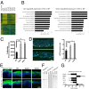
(A) RNA expression profiles compared between HaCaT WT and COSMC KO cells compared with expression profiles in human keratinocytes (Kera) and squamous skin carcinoma cells (SCC1 to -3) (for a complete list, see SI Appendix, Table S3). Genes involved in mitosis (GO:0007067) are indicated by red lines, and genes involved in epidermal development (GO:0008544) are indicated by green lines. (B) Up-regulated and down-regulated gene classes in COSMC knockout cells vs. wild-type HaCaT cells grown under conventional conditions. Epithelial cells lacking elongated O-linked glycans down-regulate genes involved in keratinocyte development and up-regulate genes involved in proliferation. (C) Proliferation assay showing 2.5-fold increased proliferation of COSMC KO#1 and KO#2 compared with HaCaT WT (n = 5). (D) IHC of organotypic skin equivalent cultures with the Ki-67 proliferation marker demonstrate enhanced proliferation and altered topology of the proliferation layer. Ki-67–positive fraction of DAPI-positive cells in COSMC KO shown at the Right (n = 3). (Scale bar: 30 μm.) (E) IHC demonstrates down-regulation of keratin 10 and dispersion of involucrin. (Scale bar: 50 μm.) (F) SDS/PAGE Western blotting of keratin 10 and unchanged keratin 5 expression (n = 3). (G) Expression profiles showed down-regulation of Notch, p53, IRF, and Foxo-3 with simultaneous up-regulation of IKBKG: an expression pattern associated with lack of differentiation and stem cell-like properties (error bars show 1× SD of replicates; *P < 0.05, **P < 0.01; NS, nonsignificant).
Fig. 4.
Fig. 4.
O-proteomics and quantitative phosphoproteomics of COSMC knockout cells. (A) The O-GalNAc proteomics strategy. GalNAc-glycopeptides are isolated on lectin weak-affinity chromatography (LWAC) and separated by IEF before nanoflow liquid chromatography tandem mass spectrometry (nLC-MS/MS) analysis. We identified 446 O-glycoproteins and 1,471 O-glycosites, of which only 369 were previously annotated in UniProt and O-GLYCBASE. (B) Global gene ontology (GO) analysis identified cellular components that carry O-linked GalNAc, including cell-adhesion proteins. Comparison was made with the total human proteome and expressed as a percentage. (C) Quantitative phosphoproteomics of COSMC KO cells versus WT. Experimental design of MS-based quantitative phosphoproteomics analysis by SILAC. Three populations of cells were labeled with normal and stable isotope-substituted arginine and lysine, creating proteins and peptides distinguishable by mass. (D) Global gene ontology (GO) analysis of the regulated set of phosphoproteins identified adhesive proteins, RAS signaling, epithelial development, and differentiation as enriched terms. Red bars represent GO terms overrepresented in WT HaCaT cells whereas blue bars represent GO terms overrepresented in KO HaCaT cells. log2 transformation of P value is indicated. (E) Illustrative examples of high-resolution MS spectrum are shown: Erk2, CD44, integrin-β4 with an increase, and ZO-1 with a decrease in phosphorylation at selective sites in mutant compared with wild-type cells. For integrin-β4, we also show an unmodified peptide with no regulation between the three conditions.
Fig. 5.
Fig. 5.
Truncation of O-linked glycans down-regulates intercellular adhesion. (A) COSMC KO cells migrated as noncoherent single cells compared with wild-type HaCaT cells in contact-inhibited monolayer cultures after scratch wound (n = 3). (B) Transmission electron microscopy micrographs of HaCaT wild-type and COSMC KO organotypic cultures. Overview of all cell layers is shown Left. Increased magnification with illustration of 5–8 cells is shown Center. (Right) Increased magnification of cell–cell interface. Increased intercellular space is noted between COSMC mutant cells (asterisks) whereas tight interaction is seen between wild-type cells (marked with arrowheads) (CM, cell membrane; NU, nucleus). (C) Cell-dissociation studies confirmed lack of cell–cell adhesion in COSMC KO HaCaT cells. Epithelial sheets of HaCaT WT cells resisted mechanical stress whereas COSMC KO HaCaT cells did not (n = 3) (***P < 0.001). (D) Keratin retraction (green) indicative of loss of desmoglein function in COSMC KO HaCaT cells. E-cadherin staining (red), which was only mildly affected, served to delineate cell membranes (n = 2). Keratin retraction was quantified by measuring the distance between the mass of the keratin bundles in areas of similar cell density as described in ref. (**P < 0.01). (Scale bar: 20 μm.) (E) Number of WT and COSMC KO cells in the cell-cycle phases (G1/0, G2, and S phase), when grown under low and high confluence. No change was observed in the number of COSMC KO cells in G1/0, G2, and S phase under high vs. low confluence, suggesting compromised contact inhibition (n = 3) (**P < 0.01). (F) p38 MAPK activation was observed in COSMC KO cells compared with wild-type cells. (G) Loss of cell adhesion in wild-type HaCaT cells treated with anisomycin (80 µg/mL) was prevented by SB203580 as detected by dispase-based dissociation assays (n = 3) (P < 0.001; anisomycin plus p38 inhibitor vs. anisomycin). (H) Loss of cell adhesion in COSMC KO was prevented by SB203580 as detected by dispase-based dissociation assays (n = 3) (*P < 0.05; **P < 0.01; DMSO vs. p38 inhibitor).

Comment in

  • Not so sweet malignant transformation.
    Baenziger JU. Baenziger JU. Proc Natl Acad Sci U S A. 2014 Sep 30;111(39):14009-10. doi: 10.1073/pnas.1415576111. Epub 2014 Sep 16. Proc Natl Acad Sci U S A. 2014. PMID: 25228771 Free PMC article. No abstract available.

References

    1. Hakomori S. Tumor-associated carbohydrate antigens defining tumor malignancy: Basis for development of anti-cancer vaccines. Adv Exp Med Biol. 2001;491:369–402. - PubMed
    1. Springer GFT. T and Tn, general carcinoma autoantigens. Science. 1984;224(4654):1198–1206. - PubMed
    1. Itzkowitz S, et al. Expression of Tn, sialosyl Tn, and T antigens in human pancreas. Gastroenterology. 1991;100(6):1691–1700. - PubMed
    1. Tarp MA, Clausen H. Mucin-type O-glycosylation and its potential use in drug and vaccine development. Biochim Biophys Acta. 2008;1780(3):546–563. - PubMed
    1. Ching CK, Holmes SW, Holmes GK, Long RG. Comparison of two sialosyl-Tn binding monoclonal antibodies (MLS102 and B72.3) in detecting pancreatic cancer. Gut. 1993;34(12):1722–1725. - PMC - PubMed

Publication types

MeSH terms