. 2014 Aug 12;7(338):ra76.
doi: 10.1126/scisignal.2005347.

Type IV collagen is an activating ligand for the adhesion G protein-coupled receptor GPR126

Affiliations

Type IV collagen is an activating ligand for the adhesion G protein-coupled receptor GPR126

Kevin J Paavola et al. Sci Signal. .

Abstract

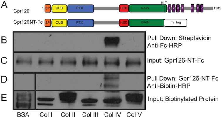
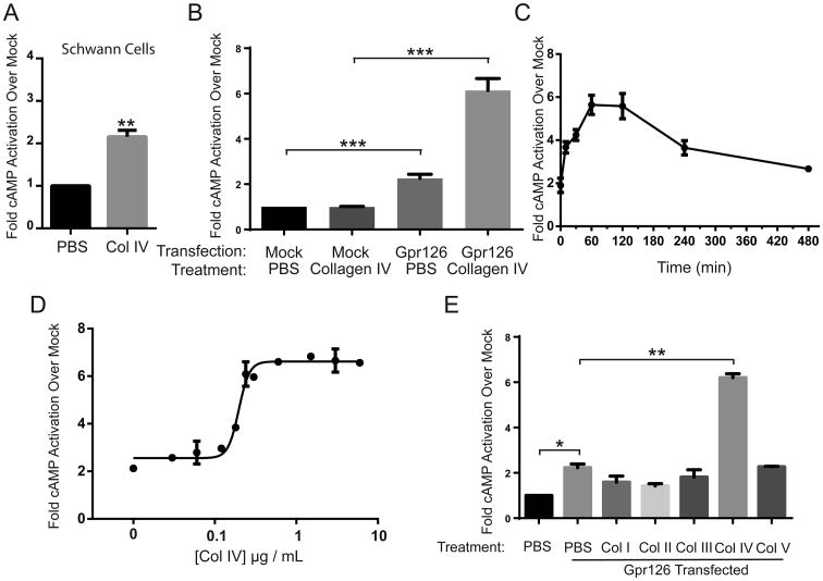
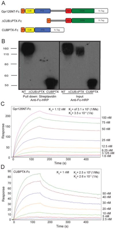
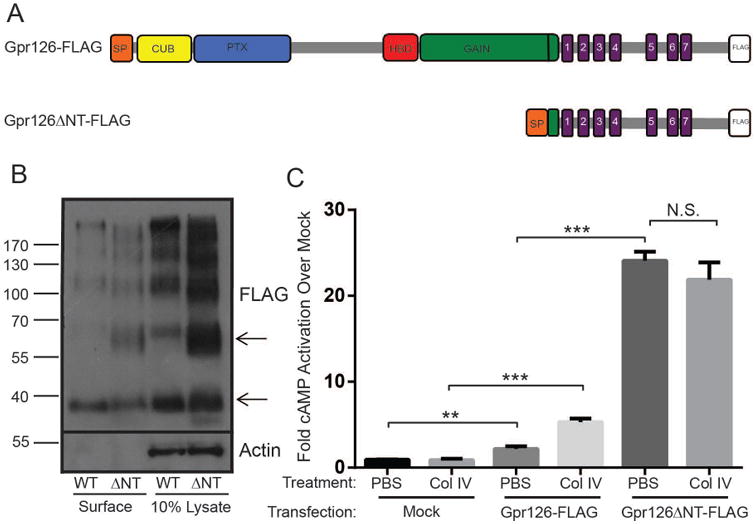
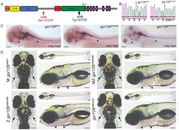
GPR126 is an orphan heterotrimeric guanine nucleotide-binding protein (G protein)-coupled receptor (GPCR) that is essential for the development of diverse organs. We found that type IV collagen, a major constituent of the basement membrane, binds to Gpr126 and activates its signaling function. Type IV collagen stimulated the production of cyclic adenosine monophosphate in rodent Schwann cells, which require Gpr126 activity to differentiate, and in human embryonic kidney (HEK) 293 cells expressing exogenous Gpr126. Type IV collagen specifically bound to the extracellular amino-terminal region of Gpr126 containing the CUB (complement, Uegf, Bmp1) and pentraxin domains. Gpr126 derivatives lacking the entire amino-terminal region were constitutively active, suggesting that this region inhibits signaling and that ligand binding relieves this inhibition to stimulate receptor activity. A new zebrafish mutation that truncates Gpr126 after the CUB and pentraxin domains disrupted development of peripheral nerves and the inner ear. Thus, our findings identify type IV collagen as an activating ligand for GPR126, define its mechanism of activation, and highlight a previously unrecognized signaling function of type IV collagen in basement membranes.


Conflict of interest statement

Competing interests: The authors declare that they have no competing interests.

Figures

Figure 1
Figure 1. Type IV collagen binds specifically to the N-terminal region of Gpr126
(A) Schematic diagram showing the conserved domains of Gpr126 including signal peptide (SP), CUB, pentraxin (PTX), hormone binding domain (HBD), GPCR autoproteolysis-inducing domain (GAIN) and seven transmembrane (7TM) region in the wild-type protein and the corresponding N-terminal fusion protein Gpr126NT-Fc. The amino acids mediating the autocatalytic cleavage of the GAIN domain are noted (HLT) and the position of the cleavage indicated by the line in the GAIN domain. (B and C) Conditioned media containing Gpr126NT-Fc fusion protein was mixed with biotinylated collagen or BSA and incubated with streptavidin agarose. Interactions (B) were detected by immunoblot using an antibody against Fc (Anti-Fc-HRP) and total input (C) was detected as a control. (D to E) In the reciprocal pull-down experiments, biotinylated collagen or BSA protein was mixed with Gpr126NT-Fc fusion protein in conditioned media and incubated with Protein A agarose. As in (B) and (C), interactions and total input were detected by immunoblot using an antibody against biotin. Blots are representative of 3 experiments.
Figure 2
Figure 2. Type IV collagen stimulates cAMP signaling in cells expressing Gpr126
(A) cAMP concentration in rat Schwann cells treated with PBS or of type IV collagen (3 μg/mL) for 1 hour. Data are mean fold change ± S.E.M. from 3 experiments. **P = 0.0013. (B) cAMP concentration in HEK293 cells transfected with empty vector or Gpr126 and treated with PBS or type IV collagen (3 μg/mL) for 1 hour. Data are mean fold change ± S.E.M. from 5 experiments. ***P = 0.0004,***P = 0.0001. (C) cAMP concentration in Gpr126-transfected HEK293 cells treated with type IV collagen (3 μg/mL) for up to 480 min. Data are mean fold change ± S.E.M. from two experiments. (D) Fold cAMP concentration in Gpr126-transfected HEK293 cells treated for 1 hour with increasing doses of type IV collagen. Data are mean fold change ± S.E.M. from two experiments. EC50 value, 0.2 μg/mL (E) cAMP concentration in HEK293 cells transfected with Gpr126 treated for 1 hour with PBS or collagen I, II, III, IV, or V (3 μg/mL) for 1 hour. Data are mean fold change ± S.E.M. from 3 experiments. **P =0.0035.
Figure 3
Figure 3. A region of Gpr126 containing CUB and Pentraxin domains mediates the high affinity interaction with type IV collagen
(A) Schematic diagram depicting truncated versions of the Gpr126NT-Fc fusion protein either containing (CUBPTX-Fc) or lacking (ΔCUBΔPTX-Fc) the CUB and PTX domains. (B) Pull down experiments using conditioned media containing Gpr126NT-Fc, ΔCUBΔPTX-Fc, or CUBPTX-Fc incubated with biotinylated type IV collagen and streptavidin agarose. Blot is representative of 4 experiments. (C to D) Biacore sensograms detecting the interaction of type IV collagen with Gpr126NT-Fc and CUBPTX-Fc. SPR monitored the binding of various concentrations of fusion proteins to immobilized collagen IV on a biosensor chip. The association and dissociation constants (Ka, Kd) for each trial are shown in the figure panels. Data are representative of two experiments; the values for association and dissociation constants are reported as mean ± SD in the text.
Figure 4
Figure 4. The N-terminal region of Gpr126 inhibits receptor signaling activity
(A) Schematic diagram showing the C-terminally FLAG tagged constructs representing full-length (Gpr126-FLAG) and N-terminally truncated Gpr126 (Gpr126ΔNT-FLAG). (B) Upper panel shows FLAG immunoblot analysis of surface expression and total lysate of HEK293 cells transfected with tagged full-length (WT) or N-terminally truncated (ΔNT) Gpr126. Lower panel shows control blot probed for actin, a cytoplasmic protein. Blot is representative of 2 experiments. (C) cAMP concentration in HEK293 cells transfected with empty vector, Gpr126-FLAG, or Gpr126ΔNT-FLAG and treated with PBS or type IV collagen (3 μg/mL) for 1 hour. Data are mean fold change ± S.E.M. from 5 experiments. **P = 0.0037; ***P = 0.0001; N.S., P = 0.36.
Figure 5
Figure 5. The gpr126st86 mutation truncates Gpr126 after the PTX domain and disrupts myelination and ear development
(A) Schematic representation of Gpr126 showing conserved domains and the mutations in gpr126st86 and previously characterized gpr126st49 (26). (B) Sequence chromatogram of heterozygous sibling and gpr126st86 mutant. STOP in the amino acid code marks a premature stop codon in the mutant. (C) Expression of mbp in wild-type (gpr126st86/+) and mutant (gpr126st86/st86 and gpr126st86/st49) larvae. Arrowheads mark mbp (or lack of it) in the lateral line nerve at 5 days post-fertilization. (D) Ear morphology in maternal (M) gpr126st86/+ mutants (which have wild-type morphology), maternal-zygotic (MZ) gpr126st86/st86 mutants, and zygotic (Z) gpr126st86/st86 mutants. Arrowheads mark the normal pericardium; arrows mark ears. Scale bars, 100 μm.

References

    1. Pierce KL, Premont RT, Lefkowitz RJ. Seven-transmembrane receptors. Nat Rev Mol Cell Biol. 2002;3:639–650. - PubMed
    1. Gudermann T, Nurnberg B, Schultz G. Receptors and G proteins as primary components of transmembrane signal transduction. Part 1. G-protein-coupled receptors: structure and function. J Mol Med (Berl) 1995;73:51–63. - PubMed
    1. Gether U, Kobilka BK. G protein-coupled receptors. II. Mechanism of agonist activation. J Biol Chem. 1998;273:17979–17982. - PubMed
    1. Hamm HE. The many faces of G protein signaling. J Biol Chem. 1998;273:669–672. - PubMed
    1. Overington JP, Al-Lazikani B, Hopkins AL. How many drug targets are there? Nat Rev Drug Discov. 2006;5:993–996. - PubMed

Publication types

MeSH terms