Review
. 2014 Sep;14(9):611-22.
doi: 10.1038/nrc3793. Epub 2014 Aug 14.

Mechanisms of disseminated cancer cell dormancy: an awakening field

Affiliations
Review

Mechanisms of disseminated cancer cell dormancy: an awakening field

María Soledad Sosa et al. Nat Rev Cancer. 2014 Sep.

Abstract

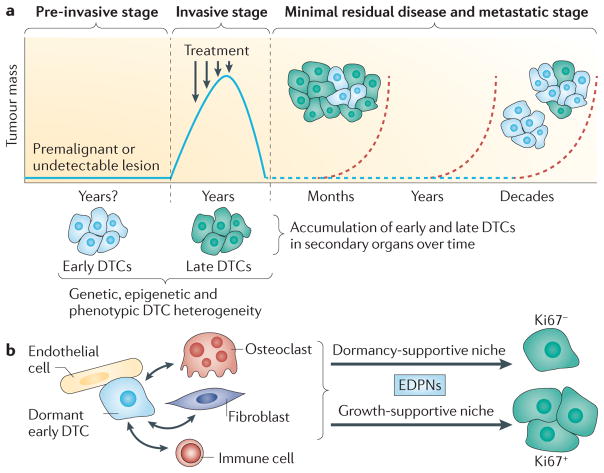
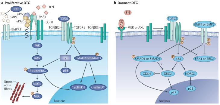
Metastases arise from residual disseminated tumour cells (DTCs). This can happen years after primary tumour treatment because residual tumour cells can enter dormancy and evade therapies. As the biology of minimal residual disease seems to diverge from that of proliferative lesions, understanding the underpinnings of this new cancer biology is key to prevent metastasis. Analysis of approximately 7 years of literature reveals a growing focus on tumour and normal stem cell quiescence, extracellular and stromal microenvironments, autophagy and epigenetics as mechanisms that dictate tumour cell dormancy. In this Review, we attempt to integrate this information and highlight both the weaknesses and the strengths in the field to provide a framework to understand and target this crucial step in cancer progression.


Conflict of interest statement

Competing interests statement

The authors declare competing interests: see Web version for details.

Figures

Figure 1
Figure 1. Dormancy of heterogeneous DTC subpopulations
a | Metastases may be initiated by and may evolve from dormant disseminated tumour cells (so-called ‘early DTCs’) from pre-invasive lesions, rather than established primary tumours,. The time required for premalignant or undectable lesions is unknown and is indicated in the figure as ‘Years?’. In fact, at the time of diagnosis, tumour cell dissemination has occurred in >50% of patients,. After primary tumour surgery and/or treatment (indicated by arrows), the tumour mass decreases and residual disease characterized by solitary dormant DTCs can be detectable for long periods (dashed blue line). After months, years or decades the metastatic tumour mass then increases (dashed red lines and tumour cell clusters). DTCs that originate from different stages of tumour evolution could form these heterogeneous masses,. For example, late DTCs, which arise from the established primary tumour may have higher metastatic potential and give rise to metastatic lesions earlier (within months of ending treatment of the primary tumour). By contrast, early DTCs (light blue) from pre-invasive lesions that remain dormant may generate metastatic tumours decades after first diagnosis. These DTCs may be the first colonizers of distant organs but may have remained in a dormant state while the primary tumour progressed. As the primary lesions progress, additional DTCs reach the target organs and contribute to dormant and proliferative DTC heterogeneity,. b | A hypothetical possibility discussed in BOX 1 is that early DTCs may, by colonizing target organs, become the founders of early DTC pre-metastatic niches (EDPNs) by crosstalk (arrows) with different host cell types such as endothelial cells, immune cells, fibroblasts and/or other stromal cells. EDPNs could support the dormancy and/or outgrowth of later arriving DTCs from later stages of evolution. Note that in part b, the same process can proceed for early DTCs that go on to form metastases independently of late DTCs (blue tumour mass in part a).
Figure 2
Figure 2. Permissive microenvironments and cues for DTC dormancy
Three non-mutually exclusive niches or microenvironments may actively support dormancy that contain active inducers of cellular dormancy and may lack signals that induce reactivation. a | Stem cell niches contain signals that also control haematopoietic stem cell (HSC) self-renewal and quiescence such as bone morphogenetic proteins (BMPs) or transforming growth factor-β2 (TGFβ2), produced by stromal cells (for example, osteoblasts). Other factors which may induce dormancy include the expression of the AXL, TGFβR3 or bone morphogenetic protein receptor type 2 (BMPR2) receptors,. In addition, this environment may also lack proliferation reactivation signals such as type I collagen-dense microenvironments, fibronectin, COCO or TGFβ3 ligand. b | The immune niche contains macrophages, CD4+ cells or CD8+ cells that may produce interferon-γ (IFNγ), which may induce dormancy in tumour cells,. In particular, tumour necrosis factor receptor 1-positive (TNFR1+) tumour cells may be susceptible to dormancy entry in this niche. c | In the vascular niche, extracellular matrix (ECM) components (for example, thrombospondin (TSP)) produced by endothelial cells not engaged in active sprouting may induce quiescence of DTCs. In addition, dormant DTCs in this niche have downregulated expression of vascular cell adhesion protein 1 (VCAM1) and lysophosphatidic acid receptors (EDG2),. The immune and vascular niches may crosstalk, as dormancy has been linked to CD4+ T cell-dependent production of CXCL9 and CXCL10, which inhibits angiogenesis (inhibitory arrow).
Figure 3
Figure 3. Intracellular pathways present in dormant and proliferative DTCs
a | In order to escape dormancy and become proliferative dormant disseminated tumour cells (DTCs) can inhibit bone morphogenetic protein (BMP) signalling by expressing inhibitors such as COCO. Proliferative DTCs also require activation of growth factor signalling via ERBB family receptors such as epidermal growth factor receptor (EGFR), which can be coupled to uPAR and β1 integrin signalling that can be activated by extracellular matrix (ECM) molecules such as fibrillar fibronectin (fFN) via α5β1 integrins or by heterotypic interaction with vascular cell adhesion protein 1 (VCAM1). Receptor tyrosine kinases like the ERBB family funnel proliferative signals through FAK, SRC and the MEK–ERK modules to cyclin D1 and cyclin D3 or through myosin light chain kinase (MLCK). Other growth-promoting pathways activated in proliferative DTCs include the canonical transforming growth factor-β1 (TGFβ1) signalling pathway. Signals between the EGFR and TGFβ signalling pathways are integrated through as yet unknown mechanisms. b | In dormant DTCs TGFβ2 and bone morphogenetic protein 4 (BMP4) or BMP7 signals predominate, activating p38, inhibiting ERK1 or ERK2 (for example, by TGFβ2, BMP7 or EDG2 inhibition), and inducing p21 and p27 cyclin-dependent kinase (CDK) inhibitors (in all cases). Specific TGFβ2 canonical (SMAD1 or SMAD5) and non-canonical signalling result in the upregulation of DEC2 and BMP7 signalling and induce NDRG1 that subsequently leads to the induction of cell cycle inhibitors and prostate cancer cell dormancy. Paradoxically, in mouse mammary cancer models NDRG1 was also shown to be induced by COCO and to support metastasis. Importantly, dormant DTCs may also be sensitive to the growth arrest-specific protein 6 (GAS6) or become unable to derive proliferative signals from fibronectin in the ECM through α5β1 integrins (previously reviewed in REF. 4). Question marks indicate unknown mechanisms or factors.

References

    1. Klein CA. Framework models of tumor dormancy from patient-derived observations. Curr Opin Genet Dev. 2010;21:42–49. - PubMed
    1. Goss PE, Chambers AF. Does tumour dormancy offer a therapeutic target? Nature Rev Cancer. 2010;10:871–877. - PubMed
    1. Klein CA. Selection and adaptation during metastatic cancer progression. Nature. 2013;501:365–372. - PubMed
    1. Aguirre-Ghiso JA. Models, mechanisms and clinical evidence for cancer dormancy. Nature Rev Cancer. 2007;7:834–846. - PMC - PubMed
    1. Demicheli R, et al. Breast cancer recurrence dynamics following adjuvant CMF is consistent with tumor dormancy and mastectomy-driven acceleration of the metastatic process. Ann Oncol. 2005;16:1449–1457. - PubMed

Publication types