. 2014 Aug 13;9(8):e103758.
doi: 10.1371/journal.pone.0103758. eCollection 2014.

Systematics of spiny predatory katydids (Tettigoniidae: Listroscelidinae) from the Brazilian Atlantic Forest based on morphology and molecular data

Affiliations

Systematics of spiny predatory katydids (Tettigoniidae: Listroscelidinae) from the Brazilian Atlantic Forest based on morphology and molecular data

Verônica Saraiva Fialho et al. PLoS One. .

Abstract

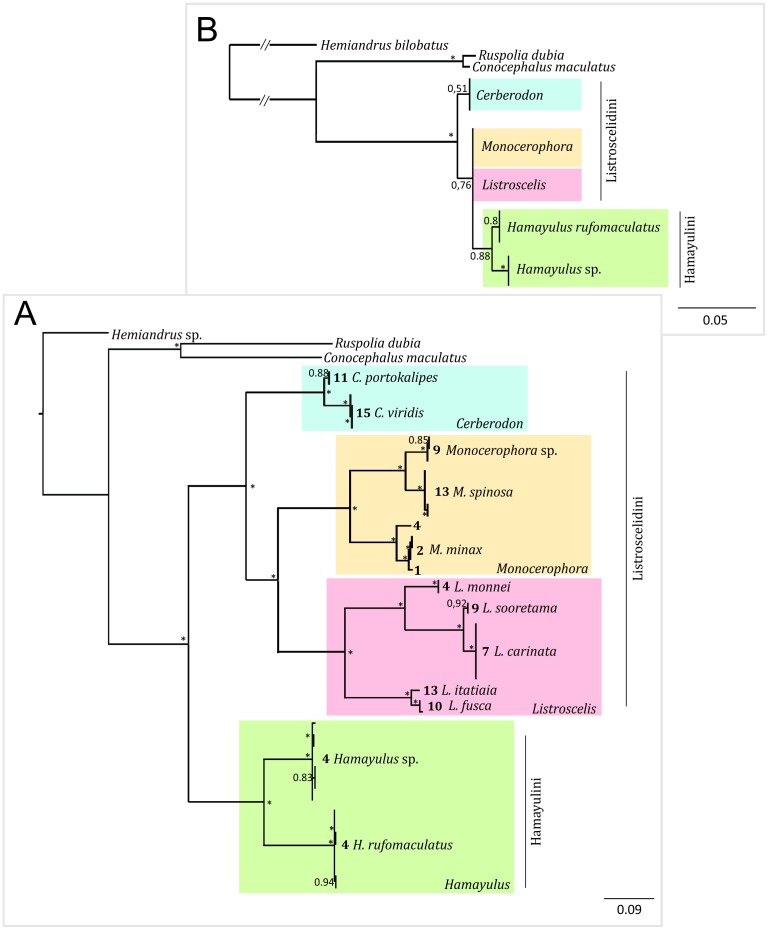
Listroscelidinae (Orthoptera: Tettigoniidae) are insectivorous Pantropical katydids whose taxonomy presents a long history of controversy, with several genera incertae sedis. This work focused on species occurring in the Brazilian Atlantic Forest, one of the world's most threatened biomes. We examined material deposited in scientific collections and visited 15 conservation units from Rio de Janeiro to southern Bahia between November 2011 and January 2012, catching 104 specimens from 10 conservation units. Based on morphological and molecular data we redefined Listroscelidini, adding a new tribe, new genus and eight new species to the subfamily. Using morphological analysis, we redescribed and added new geographic records for six species, synonymized two species and built a provisional identification key for the Atlantic Forest Listroscelidinae. Molecular results suggest two new species and a new genus to be described, possibly by the fission of the genus Hamayulus. We also proposed a 500 bp region in the final portion of the COI to be used as a molecular barcode. Our data suggest that the Atlantic Forest Listroscelidinae are seriously endangered, because they occur in highly preserved forest remnants, show high rates of endemism and have a narrow geographic distribution. Based on our results, we suggest future collection efforts must take into account the molecular barcode data to accelerate species recognition.


Conflict of interest statement

Competing Interests: The authors have declared that no competing interests exist.

Figures

Figure 1
Figure 1. Maps showing the Atlantic Forest and sampled conservation units.
Areas in light gray represent the supposed forest cover in 1500 AC (the age of Portuguese Discoveries). Areas in dark grey represent the remaining fragments of the Atlantic Forest. (A) Map of Brazil showing the AF, with the sampled area delimited by a blue rectangle (the area shown in B). (B) The 15 conservation units visited for sampling katydids (red circles). Numbers indicate the conservation units, as follows: 1 = Reserva Particular do Patrimônio Natural Serra do Teimoso, 2 = Reserva Particular do Patrimônio Natural Serra Bonita, 3 = Parque Nacional do Pau Brasil, 4 = Parque Nacional do Descobrimento, 5 = Floresta Nacional do Rio Preto, 6 = Reserva Particular do Patrimônio Natural Sítio do Zaca, 7 = Parque Estadual do Rio Doce, 8 = Estação Biológica Santa Lúcia, 9 = Reserva Biológica de Sooretama, 10 = Parque Estadual Serra do Brigadeiro, 11 = Parque Nacional do Caparaó, 12 = Parque Estadual do Ibitipoca, 13 = Parque Nacional do Itatiaia, 14 = Parque Nacional da Serra dos Órgãos, 15 = Reserva Particular do Patrimônio Natural Bacchus. Geographic coordinates of the conservation units are listed in Table S2.
Figure 2
Figure 2. Spines and tubercles on thorax and legs.
Diagrams in ventral view showing the sternum, coxae or trochanters. For each diagram, only coxa or trochanter of one leg were drawn. Spines and tubercles are indicated by light grey lines. Hamayulini trib. nov.: (A) Hamayulus rufomaculatus sp. nov., spines on sternum and mid coxae. B–D Listroscelidini: (B) Cerberodon viridis Perty, spines on sternum, mid coxae and mid trochanters, (C) Listroscelis carinata Karny, spines on sternum and spine and tubercle in mid coxae, (D) Monocerophora minax Walker, reinstated status, spines on sternum and spine and tubercle in mid coxae. Terpandrini: (E) Megatympanon speculatum Piza, spines on sternum, mid coxae and mid trochanters. All diagrams are in the same scale. Scale bars = 0.5 mm.
Figure 3
Figure 3. Gross morphology of the phallus.
Diagrams of dorsal and ventral views of the external components of the phallus. Hamayulini trib. nov.: A–B Hamayulus rufomaculatus sp. nov., (A) dorsal view, (B) ventral view. Listroscelidinae: C–D Cerberodon viridis Perty, (C) dorsal view, (D) ventral view. E–F Listroscelis magnomaculata sp. nov., (E) dorsal view, (F) ventral view. G–H Monocerophora minax Walker, reinstated status (G) dorsal view, (H) ventral view. Scale bars = 1.0 mm. Components of the phallus colored in light grey are membranous, and in dark grey sclerotized. Edges of membranous and sclerotized components devoid of membranes are in black line, and edges of portions or entire sclerotized components that are contiguous to membranes are in light grey. Abbreviations (mainly based on Snodgrass [27]): dl: dorsal lobe, e: anterior margin, low.l: lower lobe, sc: sclerite, t: titillator, up.l: upper lobe, and vl: ventral lobe.
Figure 4
Figure 4. Male postabdomen.
Diagrams in dorsal view showing the form of the epiproct, cerci, and sometimes the tergite X and paraprocts. Hamayulini trib. nov.: (A) Hamayulus rufomaculatus sp. nov.: B–K Listroscelidini: (B) Cerberodon viridis Perty, (C) Cerberodon portokalipes sp. nov., (D) Listroscelis carinata Karny, (E) Listroscelis angustifrons comb. nov., (F) Listroscelis magnomaculata sp. nov., (G) Listroscelis sooretama sp. nov., (H) Listroscelis cohni sp. nov., (I) Listroscelis fusca sp. nov., (J) Listroscelis monnei sp. nov., (K) Monocerophora minax Walker, reinstated status. Terpandrini: (L) Megatympanon speculatum Piza. Scale bars = 0.5 mm. Abbreviations (manly based on Ingrisch [28]): ce: cercus, ep: epiproct, pp: paraproct, tt: tergite X. Arrows indicate the curved spine on pp.
Figure 5
Figure 5. Male and female subgenital plates.
Diagrams in ventral view. Hamayulini trib. nov.: A–B Hamayulus rufomaculatus sp. nov., (A) male, (B) female. Listroscelidini: C–D Cerberodon viridis Perty, (C) male, (D) female. E–F Cerberodon portokalipes sp. nov., (E) male, (F) female. G–H Listroscelis carinata Karny, (G) male, (H) female. (I) Listroscelis angustifrons comb. nov., male. J–K Listroscelis magnomaculata sp. nov., (J) male, (K) female. L–M Listroscelis sooretama sp. nov., (L) male, (M) female. (N) Listroscelis cohni sp. nov., male. O–P Listroscelis fusca sp. nov., (O) male, (P) female. Q–R Listroscelis monnei sp. nov., (Q) male, (R) female. (S) Listroscelis itatiaia sp. nov., female. T–U Monocerophora minax Walker, reinstated status, (T) male, (U) female. (V) Monocerophora spinosa (Karny), female. Terpandrini: W–X Megatympanon speculatum Piza, (W) male, (X) female. Scale bars = 0.5 mm.
Figure 6
Figure 6. Stridulatory file.
Diagrams showing the arrangement of the teeth. Hamayulini trib. nov.: (A) Hamayulus rufomaculatus sp. nov. B–K Listroscelidini: (B) Cerberodon viridis Perty, (C) Cerberodon portokalipes sp. nov., (D) Listroscelis carinata Karny, (E) Listroscelis angustifrons comb. nov., (F) Listroscelis magnomaculata sp. nov., (G) Listroscelis sooretama sp. nov., (H) Listroscelis cohni sp. nov., (I) Listroscelis fusca sp. nov., (J) Listroscelis monnei sp. nov., (K) Monocerophora minax Walker, reinstated status. Terpandrini: (L) Megatympanon speculatum Piza. All diagrams are in the same scale = 0.5 mm.
Figure 7
Figure 7. Listroscelidinae Bayesian phylogenetic inferences based on COI and 18S sequences.
A) Bayesian consensus tree of 1290 aligned sites of 48 mitochondrial cytochrome oxidase I (COI) sequences (Table 3). Bold numbers on the left side of each species name correspond to the collection site of each specimen (Fig. 1). Outgroups and GenBank accession numbers (following classification in Eades et al. [14]): EU676747 = Hemiandrus sp. Ander (Stenopelmatoidea: Anostostomatidae); EF583824 = Ruspolia dubia (Redtenbacher) (Tettigonioidea: Conocephalinae: Copiphorini); NC016696 = Conocephalus maculatus (Le Guillou) (Tettigonioidea: Conocephalinae: Conocephalini). B) Simplified Bayesian consensus tree of 1615 aligned sites of 48 18S nuclear sequences. All Listroscelis and Monocerophora sequences were identical and were represented by few OTUs in the simplified tree (see the sequences numbers included in the complete tree in Table 3). Outgroups and GenBank accession numbers (following classification in Eades et al. [14]): EU676714 = Hemiandrus bilobatus Ander (Stenopelmatoidea: Anostostomatidae); JF792563 = Ruspolia dubia (Redtenbacher) (Tettigonioidea: Conocephalinae: Copiphorini); JF792565 = Conocephalus maculatus (Le Guillou) (Tettigonioidea: Conocephalinae: Conocephalini). In A and B, besides each ancestral node is a fraction number, representing its posterior probability; values >0.95 were represented by an asterisk. Listroscelidini and Hamayulini were represented in red and blue, respectively.
Figure 8
Figure 8. Habitus of Hamayulus rufomaculatus sp. nov.
A–C Holotype male, (A) lateral view, (B) dorsal view, (C) frontal view. D–F Allotype female, (D) lateral view, (E) dorsal view, (F) frontal view. Scale bars for dorsal and lateral views (horizontal bar) and frontal views (vertical bar) = 0.5 mm.
Figure 9
Figure 9. Habitus of Cerberodon viridis Perty.
A–C Male (MNRJ) from Petrópolis, in the state of Rio de Janeiro, (A) lateral view, (B) dorsal view, (C) frontal view. D–F Female (CELC) from RPPN Bacchus, in the state of Rio de Janeiro, (D) lateral view, (E) dorsal view, (F) frontal view. Scale bars for dorsal and lateral views (horizontal bar) and frontal views (vertical bar) = 0.5 mm.
Figure 10
Figure 10. Habitus of Cerberodon portokalipes sp. nov.
A–C Holotype male, (A) lateral view, (B) dorsal view, (C) frontal view. D–F Allotype female, (D) lateral view, (E) dorsal view, (F) frontal view. Scale bars for dorsal and lateral views (horizontal bar) and frontal views (vertical bar) = 0.5 mm.
Figure 11
Figure 11. Habitus of Listroscelis carinata Karny.
A–C Male (MNRJ) from Colatina, in the state of Espírito Santo, (A) lateral view, (B) dorsal view, (C) frontal view. D–F Female (CELC) from Parque Estadual do Rio Doce, in the state of Minas Gerais, (D) lateral view, (E) dorsal view, (F) frontal view. Scale bars for dorsal and lateral views (horizontal bar) and frontal views (vertical bar) = 0.5 mm.
Figure 12
Figure 12. Habitus of Listroscelis angustifrons (Piza) comb. nov.
A–C Male (NMW) from unknown locality in the state of Espírito Santo, (A) lateral view, (B) frontal view. Scale bars = 0.5 mm.
Figure 13
Figure 13. Habitus of Listroscelis magnomaculata sp. nov.
A–C Holotype male, (A) lateral view, (B) dorsal view, (C) frontal view. D–F Allotype female, (D) lateral view, (E) dorsal view, (F) frontal view. Scale bars for dorsal and lateral views (horizontal bar) and frontal views (vertical bar) = 0.5 mm.
Figure 14
Figure 14. Habitus of Listroscelis sooretama sp. nov.
A–C Holotype male, (A) lateral view, (B) dorsal view, (C) frontal view. D–F Allotype female, (D) lateral view, (E) dorsal view, (F) frontal view. Scale bars for dorsal and lateral views (horizontal bar) and frontal views (vertical bar) = 0.5 mm.
Figure 15
Figure 15. Habitus of Listroscelis cohni sp. nov.
A–C Holotype male, (A) lateral view, (B) dorsal view, (C) frontal view. Body images in the same scale = 0.5 mm.
Figure 16
Figure 16. Habitus of Listroscelis fusca sp. nov.
A–C Holotype male, (A) lateral view, (B) dorsal view, (C) frontal view. D–F Allotype female, (D) lateral view, (E) dorsal view, (F) frontal view. Scale bars for dorsal and lateral views (horizontal bar) and frontal views (vertical bar) = 0.5 mm.
Figure 17
Figure 17. Habitus of Listroselis monnei sp. nov.
A–C Holotype male, (A) lateral view, (B) dorsal view, (C) frontal view. D–F Allotype female, (D) lateral view, (E) dorsal view, (F) frontal view. Scale bars for dorsal and lateral views (horizontal bar) and frontal views (vertical bar) = 0.5 mm.
Figure 18
Figure 18. Habitus of Listroscelis itatiaia sp. nov.
A–C Holotype female, (A) lateral view, (B) dorsal view, (C) frontal view. Scale bars = 0.5 mm.
Figure 19
Figure 19. Habitus of Monocerophora minax Walker, reinstated status.
A–C Male (CELC) from RPPN Serra do Teimoso, in the state of Bahia, (A) lateral view, (B) dorsal view, (C) frontal view. D–F Female (CELC) from RPPN Serra do Teimoso, in the state of Bahia, (D) lateral view, (E) dorsal view, (F) frontal view. Scale bars for dorsal and lateral views (horizontal bar) and frontal views (vertical bar) = 0.5 mm.
Figure 20
Figure 20. Habitus of Monocerophora spinosa (Karny).
A–C Holotype male (NMW), (A) lateral view, (B) dorsal view, (C) frontal view. D–F Female (CELC) from Parque Nacional do Itatiaia, in the state of Rio de Janeiro, (D) lateral view, (E) dorsal view, (F) frontal view. Scale bars for dorsal and lateral views (horizontal bar) and frontal views (vertical bar) = 0.5 mm.
Figure 21
Figure 21. Habitus of Megatympanon speculatum Piza.
A–C Male (MNRJ) from Petrópolis, in the state of Rio de Janeiro, (A) lateral view, (B) dorsal view, (C) frontal view. D–F Female (MNRJ) from Itaguaí, in the state of Rio de janeiro, (D) lateral view, (E) dorsal view, (F) frontal view. Scale bars for dorsal and lateral views (horizontal bar) and frontal views (vertical bar) = 0.5 mm.
Figure 22
Figure 22. Geographic distribution of the species of Listroscelidinae in the Brazilian Atlantic Forest.
(A) Registers of Hamayulus rufomaculatus sp. nov. (red square), Hamayulus sp. (blue inverted triangle) and Megatympanon speculatum Piza (green circle). (B) Registers of Monocerophora minax Walker, reinstated status (yellow plus), M. spinosa (Karny) (purple plus), Monocerophora sp. (green plus), Cerberodon portokalipes sp. nov. (dark red square) and C. viridis Perty (blue square). (C) Registers of Listroscelis angustifrons (Piza) (inverted green triangle), L. atrata Redtenbacher (blue square), L. carinata Karny (purple star), L. cohni sp. nov. (orange circle) and L. ferruginea Redtenbacher (red triangle). (D) Registers of Listroscelis fusca sp. nov. (yellow star), L. itatiaia sp. nov. (inverted red triangle), L. magnomaculata sp. nov. (purple square), L. monnei sp. nov. (dark blue circle) and L. sooretama sp. nov. (green triangle).

Similar articles

Cited by

References

    1. Bruner L (1915) Notes on tropical American Tettigonoidea (Locustodea). Ann Carnegie Mus 9: 284–404.
    1. Redtenbacher J (1891) Monographie der Conocephaliden. Verh Zool Bot Ges Wien 41: 315–562.
    1. Saussure deH, Pictet A (1898) Insecta Orthoptera (Orthoptera Genuina). Fam. Locustidae. In: Goodman FD, Salvin O, editors. Biologia Centrali-Americana. v. 1. pp. 345–456.
    1. Karny H (1911) Descriptions Conocephalidarum novarum. Verh Zool Bot Ges Wien 61: 334–347.
    1. Kirby WF (1906) A Synonymic Catalogue of Orthoptera (Orthoptera Saltatoria, Locustidae vel Acridiidae), Vol. 2. London: British Museum (Natural History). 562 p.

Publication types

Associated data

LinkOut - more resources