Review
. 2014 Sep;15(9):565-76.
doi: 10.1038/nrm3854. Epub 2014 Aug 13.

Regulation of microRNA function in somatic stem cell proliferation and differentiation

Affiliations
Review

Regulation of microRNA function in somatic stem cell proliferation and differentiation

Archana Shenoy et al. Nat Rev Mol Cell Biol. 2014 Sep.

Abstract

microRNAs (miRNAs) are important modulators of development. Owing to their ability to simultaneously silence hundreds of target genes, they have key roles in large-scale transcriptomic changes that occur during cell fate transitions. In somatic stem and progenitor cells--such as those involved in myogenesis, haematopoiesis, skin and neural development--miRNA function is carefully regulated to promote and stabilize cell fate choice. miRNAs are integrated within networks that form both positive and negative feedback loops. Their function is regulated at multiple levels, including transcription, biogenesis, stability, availability and/or number of target sites, as well as their cooperation with other miRNAs and RNA-binding proteins. Together, these regulatory mechanisms result in a refined molecular response that enables proper cellular differentiation and function.


Figures
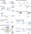
Figure 1
Figure 1. Regulation of miRNA biogenesis and function occurs at multiple levels
a | microRNA (miRNA) expression is regulated by transcriptional and epigenetic activators and repressors. b | Post-transcriptional regulation can be modulated at the level of processing. A complex composed of DGCR8 and Drosha cleaves the primary transcript to generate a precursor miRNA hairpin, which is then processed by Dicer to generate the mature product. miR-451 is unique as it bypasses Dicer processing and is directly processed by argonaute 2 (AGO2). c | Mature miRNAs and their processing intermediates (pre-miRNAs) can be destabilized; for example, members of the let-7 miRNA family, which are destabilized by the RNA-binding protein (RBP) LIN28A. d | Multiple miRNAs can act on a single mRNA cooperating to destabilize the transcript. e | A single miRNA can target many mRNAs in a gene network that drives a specific cell fate transition. f | Alternative mRNA polyadenylation results in 3′ untranslated regions (3′ UTRs) of different lengths, which therefore may or may not contain miRNA target sites. g | RBPs can bind an mRNA and promote or inhibit miRNA activity at a neighbouring site, leading to mRNA degradation or translation, respectively. h | Expression of endogenous ‘sponges’ (with sequences partially complementary to miRNAs) has been proposed to interfere with miRNA-mediated inhibition of a target mRNA, thus promoting messenger stability and translation. PAS, polyadenylation site.
Figure 2
Figure 2. Lineage-restricted stem and progenitor cell differentiation during the development of four model lineages
a | In skeletal muscle, progenitors that give rise to myoblasts arise from the paraxial mesoderm. Myoblasts terminally differentiate into muscle cells, which fuse to give rise to myotubes. The myotubes can produce fast-twitch or slow-twitch muscle fibres. Muscle progenitors also give rise to satellite cells, which serve as adult stem cells of the muscle. b | In the blood, self-renewing haematopoietic stem cells (HSCs) give rise to multipotent progenitors (MPPs), which further differentiate into the progenitors of lymphoid lineage (common lymphoid progenitor (CLP)) or myeloid lineage (common myeloid progenitor (CMP)). CLPs differentiate into progenitors of B cells (BCPs) and the progenitors of T cells and natural killer cells (TNKs). CMPs give rise to megakaryocyte–erythroid progenitors (MEPs; which differentiate into erythrocytes and platelets) and the granulocyte–macrophage progenitors (GMPs; which differentiate into monocytes, neutrophils and eosinophils). c | The epidermis is a stratified epithelium composed of progenitor cells in the innermost basal layer and successive layers of differentiating cells as one moves to the surface. The hair follicle develops when primitive epidermal cells form placodes that differentiate into hair follicle stem cells, which in turn undergo further sequential differentiation to produce the outer root sheath (ORS) progenitor cells, the matrix cells, the inner root sheath (IRS) cells, and finally the mature hair follicle. Epithelial stem cells at the junction of the epidermis and hair follicle give rise to the sebaceous gland. d | Neural — neuroepithelial cells expand and produce early neurons, as well as radial glial cells (RGCs). For most of embryogenesis, RGCs divide asymmetrically producing a neuron and a new RGC. During the end of embryogenesis and in early postnatal stages, RGCs acquire gliogenic competence (astrocyte precursor cell (APC) and oligodendrocyte progenitor cell (OPC) for the astrocyte and oligodendrocyte precursor cell, respectively), giving rise to astrocytes and oligodendrocytes. In later postnatal stages, adult neural stem cells (aNSCs) can give rise to neurons or glia. IFE, interfollicular epidermis. HSPCs, haematopoietic stem and progenitor cells.
Figure 3
Figure 3. miRNA regulation during myogenesis
a | microRNAs (miRNAs) that are expressed at high levels in myoblasts (light green box) promote cell cycle progression by suppressing the translation of the cell cycle inhibitor p27. miRNAs display increased expression during myogenesis (dark green box) and are transcriptionally activated by myogenic transcription factors such as MYF5, MyoD and myogenin. These miRNAs both repress genes that promote the progenitor fate and repress genes that silence the muscle programme. For example, some miRNAs inhibit the transcription factors PAX3 and PAX7 and repress the cell cycle regulator CDC25A. Others inhibit the epigenetic repressor histone deacetylase 4 (HDAC4), which in turn leads to derepression of the muscle transcription factor MEF2 and subsequent upregulation of muscle genes. b | Regulation of miRNA activity during myogenesis. miRNA activity during muscle differentiation is modulated by the RNA-binding protein HuR, which binds next to the miR-1192 binding site on the 3′ untranslated region (3′ UTR) of the transcript encoding the chromatin-binding protein HMGB1, inhibiting miRNA-mediated suppression of this gene. Alternative polyadenylation of the mRNA encoding PAX3 results in either a long form that contains a binding site for miR-206 in its 3′ UTR or a short form that does not contain the binding site and, therefore, is resistant to miRNA-mediated suppression. H19 is a long non-coding RNA with multiple let-7 binding sites, which has been proposed to titrate let-7 away from other targets such as Dicer and HMGA2, thus relieving the suppression of these targets. Dark lines represent activation and grey bars represent suppression.
Figure 4
Figure 4. miRNAs regulate cell fate choices and differentiation during haematopoiesis
a | microRNAs (miRNAs) enriched in one cell type (outlined by the colour of the cell type) can promote the differentiation of progenitors (miR-223) or can inhibit the alternative lineages of oligopotent progenitors (miR-9 and miR-150). miRNAs also inhibit apoptosis in progenitors during haematopoiesis (miR-125 targets pro-apoptotic genes BAK1, KLF13 and BMF, and miR-17-92 targets BIM). The inhibition of miRNAs (miR-125/351) by a KRAB-ZFP transcription factor and its co-repressor KAP1 is necessary to enable mitophagy during erythrocyte maturation. miRNA levels are regulated at the transcriptional level. Levels of miR-223 are modulated by sequential binding of the transcription factors NF1A and CCAAT/enhancer-binding protein-β (CEBP). Binding of CEBP leads to maximal activation in granulocytes. The miR-451 locus is activated by binding of the erythrocyte-specific transcription factor GATA1. b | miRNAs that regulate developmental transitions in haematopoiesis from the embryo to the adult are indicated. The modulation of let-7 expression leads to a reduction in self-renewal in adult haematopoietic stem cells (HSCs) relative to fetal HSCs. In fetal HSCs, high levels of RNA-binding protein LIN28B repress let-7 biogenesis, which in turn leads to high levels of HMGA2. In adult HSCs, LIN28B levels are low, which leads to high let-7 activity and repression of the chromatin factor HMGA2. c | miRNAs expressed in niche cells can regulate stem cell proliferation and differentiation. For example, miR-126, expressed in neighbouring mesenchyme cells (outlined by a lilac box), represses the adhesion molecule vascular cell adhesion protein 1 (VCAM1) to promote maturation of primitive HSCs. CLP, common lymphoid progenitor; CMP, common myeloid progenitor; GMP, granulocyte–monocyte progenitor; MEP, megakaryocyte–erythroid progenitor. HSPC, haematopoietic stem and progenitor cell.
Figure 5
Figure 5. miRNAs regulate the development of the epidermis and hair follicle
microRNAs (miRNAs) function during differentiation of epidermal stem cells into cells of the interfollicular epidermis (IFE). miR-205 and miR-125 promote self-renewal of epidermal stem cells but are downregulated during differentiation. miR-205 suppresses multiple inhibitors of the PI3K–AKT pathway, thereby promoting AKT signalling. By contrast, miR-203, upregulated during differentiation, targets genes that encode proteins that promote cell cycle progression (SKP2, p63, MSI2 and VAV3) thereby inducing cell cycle exit. miRNAs highly expressed in stem cells and differentiated progeny are outlined in orange and red, respectively.
Figure 6
Figure 6. miRNAs regulate the development of neural cells
a | microRNAs (miRNAs) that promote neurogenesis are often integrated into positive transcriptional and epigenetic feedback loops. The transcription factor TLX recruits the histone H3K4 demethylase LSD1 to inhibit miR-9 and miR-137 in neural stem cells (NSCs). During differentiation, miR-9 and miR-137 levels increase and target TLX and LSD1, respectively. Similarly, the methylated DNA binding protein MBD1 inhibits miR-195 in NSCs. During differentiation, miR-195 levels increase and this represses MBD1. Additionally, the REST complex, which represses neuronal gene expression, is highly expressed in NSCs and represses miR-9 and miR-124. During differentiation, REST levels decrease and miR-9 and miR-124 levels increase. miR-9 and miR-124 target components of the REST complex during differentiation. b | miRNAs enriched in oligodendrocytes (dark blue) promote oligodendrocyte differentiation by targeting transcription factors that are highly expressed in progenitors (HES5, SOX6, FOXJ3 and ZFP238) and signalling pathway components (platelet-derived growth factor receptor A (PDGFRA)). miRNAs upregulated during neuronal differentiation (let-7, miR-124 and miR-9*) (dark blue) promote cell cycle exit by inhibiting the cell cycle regulator cyclin D, chromatin remodeller BAF53A and transcription factors (such as SOX9) that are important for the progenitor state. c | miRNAs upregulated during neuronal differentiation (dark blue) promote derepression of neuronal gene expression (miR-26 and miR-124 targeting the REST complex, and miR-9/let-7 targeting TLX) and neuron-specific splicing (miR-124 targeting splicing regulator polypyrimidine tract-binding protein 1 (PTBP1)). miR-9* and miR-124 also promote the formation of a neuron-specific chromatin remodelling complex by inhibiting a progenitor-specific subunit (miR-9* and miR-124 targeting BAF53A) of the complex. OPC, oligodendrocyte progenitor cell.

Similar articles

Cited by

References

    1. Ha M, Kim VN. Regulation of microRNA biogenesis. Nature Rev. GMo. Cell Biol. 2014;15:509–524. - PubMed
    1. Babiarz JE, Blelloch R. Small RNAs – their biogenesis, regulation and function in embryonic stem cells. StemBook. 2009 - PubMed
    1. Bernstein E, et al. Dicer is essential for mouse development. Nature Genet. 2003;35:215–217. - PubMed
    1. Wang Y, Medvid R, Melton C, Jaenisch R, Blelloch R. DGCR8 is essential for microRNA biogenesis and silencing of embryonic stem cell self-renewal. Nature Genet. 2007;39:380–385. - PMC - PubMed
    1. Huntzinger E, Izaurralde E. Gene silencing by microRNAs: contributions of translational repression and mRNA decay. Nature Rev. Genet. 2011;12:99–110. - PubMed

Publication types