. 2014 Aug 14;5(8):e1375.
doi: 10.1038/cddis.2014.338.

Impaired cell death and mammary gland involution in the absence of Dock1 and Rac1 signaling

Affiliations

Impaired cell death and mammary gland involution in the absence of Dock1 and Rac1 signaling

H Bagci et al. Cell Death Dis. .

Abstract

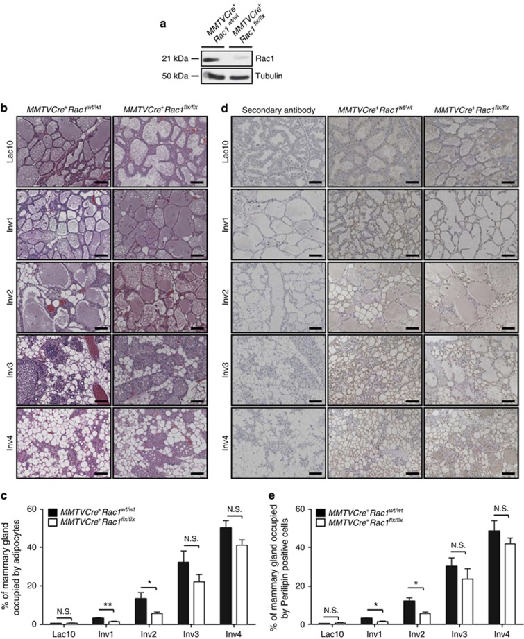
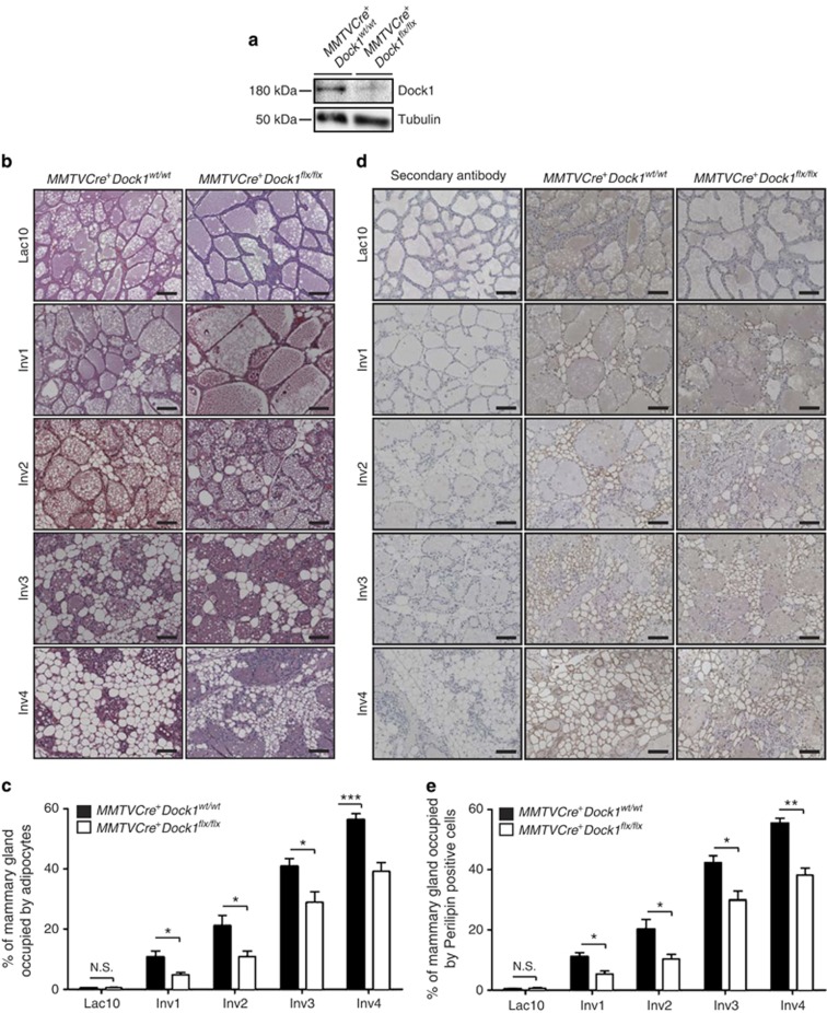
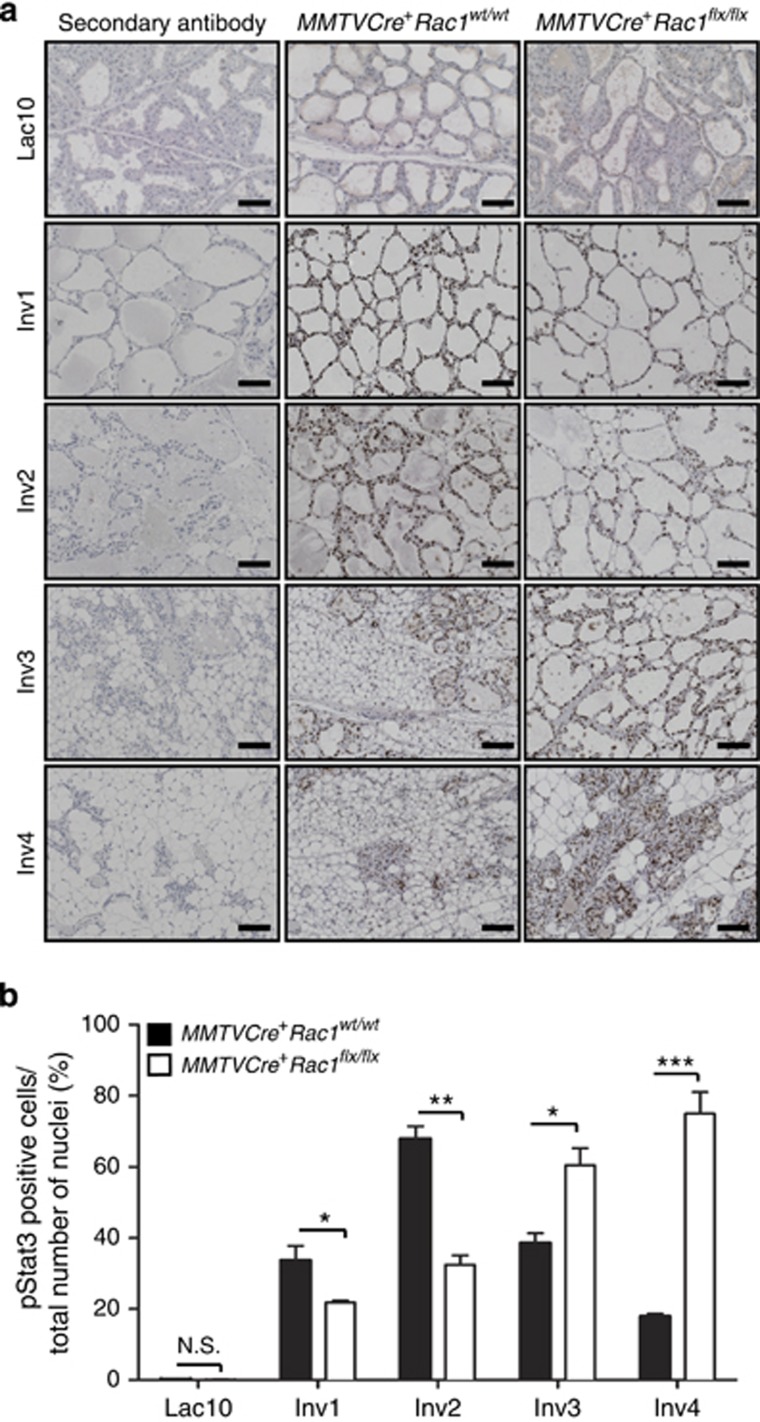
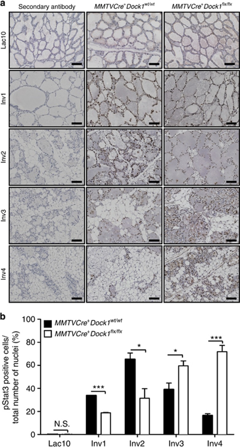
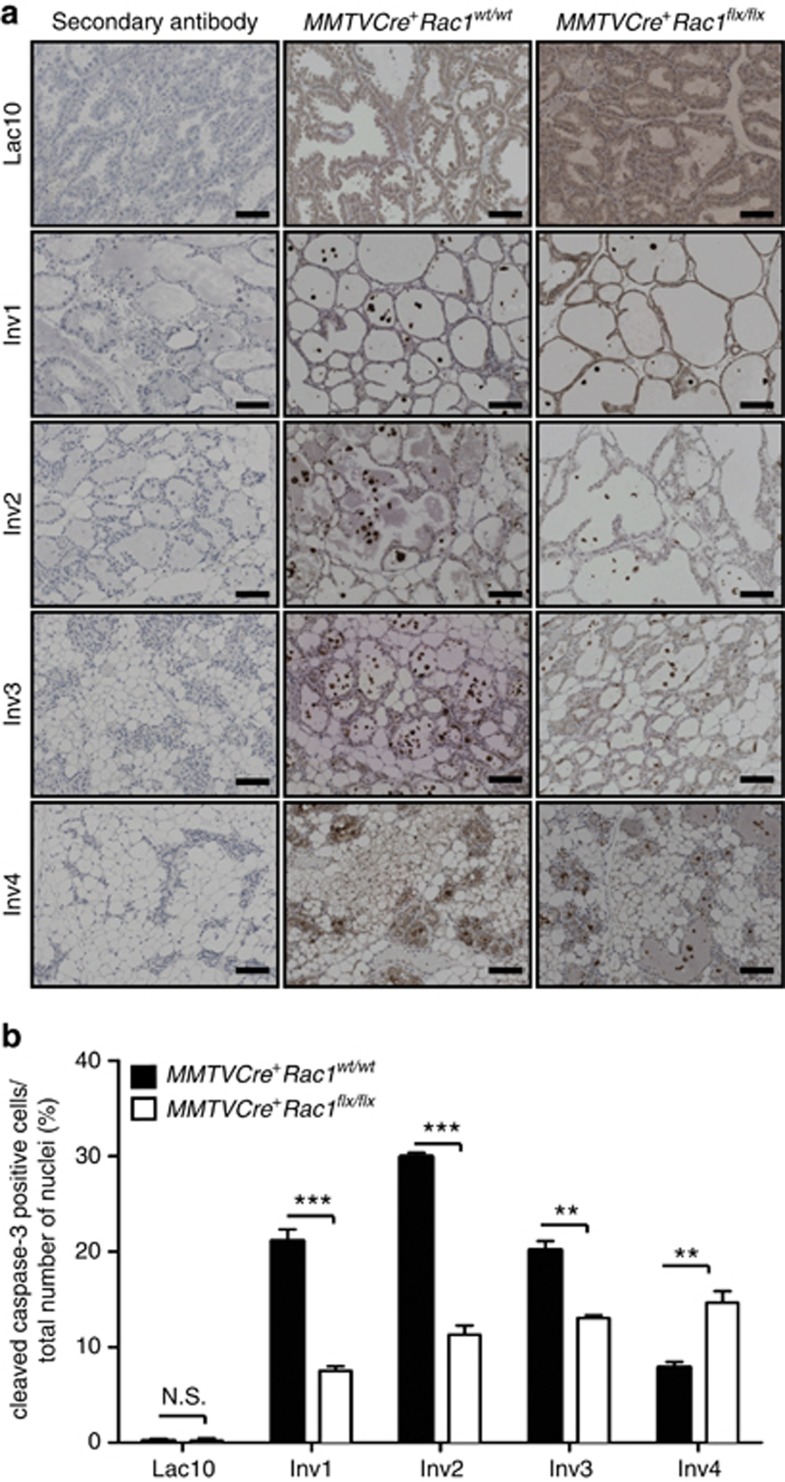
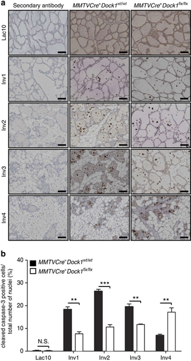
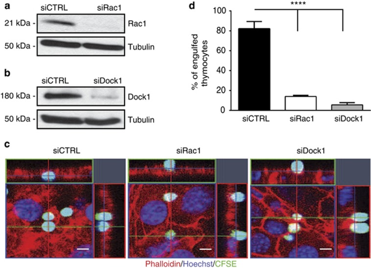
Throughout life, the tight equilibrium between cell death and the prompt clearance of dead corpses is required to maintain a proper tissue homeostasis and prevent inflammation. Following lactation, mammary gland involution is triggered and results in the death of excessive epithelial cells that are rapidly cleared by phagocytes to ensure that the gland returns to its prepregnant state. Orthologs of Dock1 (dedicator of cytokinesis 1), Elmo and Rac1 (ras-related C3 botulinum toxin substrate 1) in Caenorhabditis elegans are part of a signaling module in phagocytes that is linking apoptotic cell recognition to cytoskeletal reorganization required for engulfment. In mammals, Elmo1 was shown to interact with the phosphatidylserine receptor Bai1 and relay signals to promote phagocytosis of apoptotic cells. Still, the role of the RacGEF Dock1 in the clearance of dying cells in mammals was never directly addressed. We generated two mouse models with conditional inactivation of Dock1 and Rac1 and revealed that the expression of these genes is not essential in the mammary gland during puberty, pregnancy and lactation. We induced mammary gland involution in these mice to investigate the role of Dock1/Rac1 signaling in the engulfment of cell corpses. Unpredictably, activation of Stat3 (signal transducer and activator of transcription 3), a key regulator of mammary gland involution, was impaired in the absence of Rac1 and Dock1 expression. Likewise, failure to activate properly Stat3 was coinciding with a significant delay in the initiation and progression of mammary gland involution in mutant animals. By using an in vitro phagocytosis assay, we observed that Dock1 and Rac1 are essential to mediate engulfment in epithelial phagocytes. In vivo, cell corpses accumulated at late time points of involution in Dock1 and Rac1 mutant mammary glands. Overall, our study demonstrated an unsuspected role for Dock1/Rac1 signaling in the initiation of mammary gland involution, and also suggested a role for this pathway in the clearance of dead cells by epithelial phagocytes.


Figures

Figure 1
Figure 1
Mammary gland involution is delayed in MMTVCre+Rac1flx/flx mice. (a) Western blot analysis demonstrating the absence of Rac1 expression in Lac10 mammary glands of MMTVCre+Rac1flx/flx mice. (b) H&E staining of mammary glands at 10 days of lactation and after 1, 2, 3 or 4 days of involution showing delayed repopulation of adipocytes in MMTVCre+Rac1flx/flx mice (scale bar: 100 μm, × 20). (c) Average percentage of the area occupied by adipocytes at the indicated lactation and involution days in MMTVCre+Rac1wt/wt and MMTVCre+Rac1flx/flx mammary gland. Twenty random sections were analyzed and quantified from each mouse (n=6). Data were shown as mean±S.E.M. The indicated P-values were calculated by using a two-tailed Student's t test. NS, not significant, *P≤0.05 and **P≤0.01. (d) IHC analysis showing Perilipin staining in mammary glands at 10 days of lactation and after 1, 2, 3 and 4 days of involution showing delayed repopulation of adipocytes in MMTVCre+Rac1flx/flx mice (scale bar: 100 μm, × 20x). (e) Average percentage of the area occupied by Perilipin-positive cells at the indicated lactation and involution days in MMTVCre+Rac1wt/wt and MMTVCre+Rac1flx/flx mice. Ten random sections were analyzed and quantified from each mouse (n=3). Data were shown as mean±S.E.M. The indicated P-values were calculated by using a two-tailed Student's t test. *P≤0.05
Figure 2
Figure 2
Mammary gland involution is delayed in MMTVCre+Dock1flx/flx mice. (a) Western blot analysis demonstrating the absence of Dock1 expression in Lac10 mammary glands of MMTVCre+Dock1flx/flx mice. (b) H&E staining of mammary glands at 10 days of lactation and after 1, 2, 3 or 4 days of involution showing delayed repopulation of adipocytes in MMTVCre+Dock1flx/flx mice (scale bar: 100 μm, × 20). (c) Average percentage of the area occupied by adipocytes at the indicated lactation and involution days in MMTVCre+Dock1wt/wt and MMTVCre+Dock1flx/flx mammary gland. Twenty random sections were analyzed and quantified from each mouse (n=6). Data were shown as mean±S.E.M. The indicated P-values were calculated by using a two-tailed Student's t-test. NS, not significant, *P≤0.05 and ***P≤0.001. (d) IHC analysis showing Perilipin staining in mammary glands at 10 days of lactation and after 1, 2, 3 and 4 days of involution showing delayed repopulation of adipocytes in MMTVCre+Dock1flx/flx mice (scale bar: 100 μm, × 20). (e) Average percentage of the area occupied by Perilipin-positive at the indicated lactation and involution days in MMTVCre+Dock1wt/wt and MMTVCre+Dock1flx/flx mice. Ten random sections were analyzed and quantified from each mouse (n=3). Data were shown as mean±S.E.M. The indicated P-values were calculated by using a two-tailed Student's t-test. NS, not significant, *P≤0.05 and **P≤0.01
Figure 3
Figure 3
Stat3 activation is delayed in the absence of Rac1 expression during involution. (a) IHC analysis showing p-Stat3 staining in mammary glands at 10 days of lactation and after 1, 2, 3 and 4 days of involution in MMTVCre+Rac1wt/wt and MMTVCre+Rac1flx/flx mice (scale bar: 100 μm, × 20). (b) Average number of the p-Stat3-positive MECs normalized by the total number of nuclei (%) at the indicated lactation and involution days in MMTVCre+Rac1wt/wt and MMTVCre+Rac1flx/flx mice. Ten random sections were analyzed and quantified from each mouse (n=3). Data were shown as mean±S.E.M. The indicated P-values are calculated by using a two-tailed Student's t-test. NS, not significant, *P≤0.05, **P≤0.01 and ***P≤0.001
Figure 4
Figure 4
Stat3 activation is delayed in the absence of Dock1 expression during involution. (a) IHC analysis showing p-Stat3 staining in mammary glands at 10 days of lactation and after 1, 2, 3 and 4 days of involution in MMTVCre+Dock1wt/wt and MMTVCre+Dock1flx/flx mice (scale bar: 100 μm, × 20). (b) Average number of the p-Stat3-positive MECs normalized by the total number of nuclei (%) at the indicated lactation and involution days in MMTVCre+Dock1wt/wt and MMTVCre+Dock1flx/flx mice. Ten random sections were analyzed and quantified from each mouse (n=3). Data were shown as mean±S.E.M. The indicated P-values are calculated by using a two-tailed Student's t-test. NS, not significant, *P≤0.05 and ***P≤0.001
Figure 5
Figure 5
Cell death is decreased in the absence of Rac1 expression during involution. (a) IHC analysis showing cleaved caspase-3 staining in mammary glands at 10 days of lactation and after 1, 2, 3 and 4 days of involution in MMTVCre+Rac1wt/wt and MMTVCre+Rac1flx/flx mice (scale bar: 100 μm, × 20) (b) Average number of the cleaved caspase-3-positive MECs normalized by the total number of nuclei (%) at the indicated lactation and involution days in MMTVCre+Rac1wt/wt and MMTVCre+Rac1flx/flx mice. Ten random sections were analyzed and quantified from each mouse (n=3). Data were shown as mean±S.E.M. The indicated P-values are calculated by using a two-tailed Student's t-test. NS, not significant, **P≤0.01 and ***P≤0.001
Figure 6
Figure 6
Cell death is decreased in the absence of Dock1 expression during involution. (a) IHC analysis showing cleaved caspase-3 staining in mammary glands at 10 days of lactation and after 1, 2, 3 and 4 days of involution in MMTVCre+Dock1wt/wt and MMTVCre+Dock1flx/flx mice (scale bar: 100 μm, × 20) (b) Average number of the cleaved caspase-3-positive MECs normalized by the total number of nuclei (%) at the indicated lactation and involution days in MMTVCre+Dock1wt/wt and MMTVCre+Dock1flx/flx mice. Ten random sections were analyzed and quantified from each mouse (n=3). Data were shown as mean±S.E.M. The indicated P-values are calculated by using a two-tailed Student's t-test. NS, not significant, **P≤0.01 and ***P≤0.001
Figure 7
Figure 7
Phagocytosis of apoptotic cells by MECs requires Rac1 and Dock1 expression. (a) Western blot analysis demonstrating siRNA-mediated knockdown of Rac1 expression in NMuMG cells. (b) Western blot analysis demonstrating siRNA-mediated knockdown of Dock1 expression in NMuMG cells. (c) Representative confocal microscopy images showing defective engulfment of apoptotic thymocytes by NMuMG cells transfected with siRac1 and siDock1 (scale bar: 5 μm; red: phalloidin staining; blue: Hoechst; green: CFSE). (d) Average percentage of apoptotic thymocytes engulfed by NMuMG cells transfected with siCTRL, siRac1 and siDock1 after 2 h (n=3). Data were shown as mean±S.D. The indicated P-values are calculated by using one-way ANOVA, followed by a Bonferroni's multiple comparisons test. ****P≤0.0001

Similar articles

Cited by

References

    1. Fuchs Y, Steller H. Programmed cell death in animal development and disease. Cell. 2011;147:742–758. - PMC - PubMed
    1. Elliott MR, Ravichandran KS. Clearance of apoptotic cells: implications in health and disease. J Cell Biol. 2010;189:1059–1070. - PMC - PubMed
    1. Reddien PW, Horvitz HR. The engulfment process of programmed cell death in Caenorhabditis elegans. Annu Rev Cell Dev Biol. 2004;20:193–221. - PubMed
    1. Albert ML, Kim JI, Birge RB. alphavbeta5 integrin recruits the CrkII–Dock180–rac1 complex for phagocytosis of apoptotic cells. Nat Cell Biol. 2000;2:899–905. - PubMed
    1. Gumienny TL, Brugnera E, Tosello-Trampont AC, Kinchen JM, Haney LB, Nishiwaki K, et al. CED-12/ELMO, a novel member of the CrkII/Dock180/Rac pathway, is required for phagocytosis and cell migration. Cell. 2001;107:27–41. - PubMed

Publication types

MeSH terms