. 2014 Aug 13;9(8):e104696.
doi: 10.1371/journal.pone.0104696. eCollection 2014.

Successful non-surgical deep uterine transfer of porcine morulae after 24 hour culture in a chemically defined medium

Affiliations

Successful non-surgical deep uterine transfer of porcine morulae after 24 hour culture in a chemically defined medium

Emilio A Martinez et al. PLoS One. .

Abstract

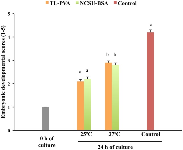
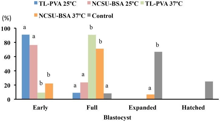
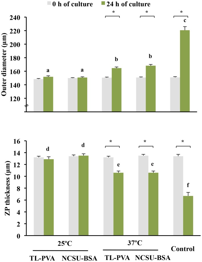
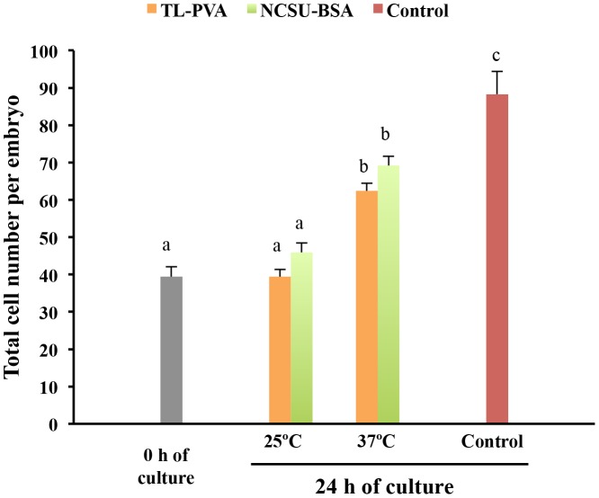
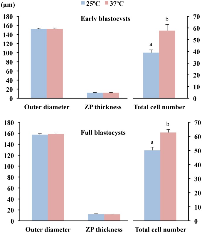
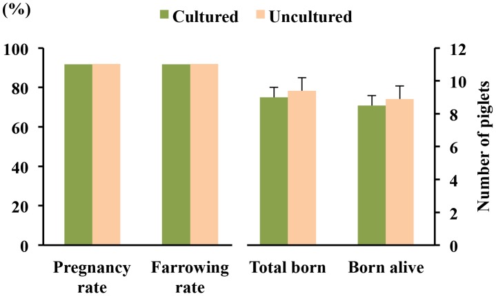
Excellent fertility and prolificacy have been reported after non-surgical deep uterine transfers of fresh in vivo-derived porcine embryos. Unfortunately, when this technology is used with vitrified embryos, the reproductive performance of recipients is low. For this reason and because the embryos must be stored until they are transferred to the recipient farms, we evaluated the potential application of non-surgical deep uterine transfers with in vivo-derived morulae cultured for 24 h in liquid stage. In Experiment 1, two temperatures (25 °C and 37 °C) and two media (one fully defined and one semi-defined) were assessed. Morulae cultured in culture medium supplemented with bovine serum albumin and fetal calf serum at 38.5 °C in 5% CO2 in air were used as controls. Irrespective of medium, the embryo viability after 24 h of culture was negatively affected (P<0.05) at 25 °C but not at 37 °C compared with the controls. Embryo development was delayed in all experimental groups compared with the control group (P<0.001). Most of the embryos (95.7%) cultured at 37 °C achieved the full or expanded blastocyst stage, and unlike the controls, none of them hatched at the end of culture. In Experiment 2, 785 morulae were cultured in the defined medium at 37 °C for 24 h, and the resulting blastocysts were transferred to the recipients (n = 24). Uncultured embryos collected at the blastocyst stage (n = 750) were directly transferred to the recipients and used as controls (n = 25). No differences in farrowing rates (91.7% and 92.0%) or litter sizes (9.0 ± 0.6 and 9.4 ± 0.8) were observed between the groups. This study demonstrated, for the first time, that high reproductive performance can be achieved after non-surgical deep uterine transfers with short-term cultured morulae in a defined medium, which opens new possibilities for the sanitary, safe national and international trade of porcine embryos and the commercial use of embryo transfer in pigs.


Conflict of interest statement

Competing Interests: The authors have the following interests: The grant from the Selección Batalle S.A. is unrestricted and not related to any employment, consultancy, patents, products in development, or marketed products, etc. Co-authors JV, IC, MD, and JR have a position at Selección Batallé S.A. This does not alter the authors' adherence to all the PLoS ONE policies on sharing data and materials.

Figures

Figure 1
Figure 1. Developmental scores (means ± S.E.M.) of in vivo-derived morulae cultured for 24 h using different temperatures and media.
Morulae were cultured in closed Eppendorf tubes containing 1.5 mL of TL-PVA or NCSU23-BSA media, supplemented with 10 mM HEPES, at 25°C or 37°C. Controls were morulae cultured in 4-well multi-dish plates, with each well containing 500 µL of NCSU-23 medium supplemented with BSA and fetal calf serum at 38.5°C in humidified air with 5% CO2. The developmental stage was scored according to the following classes: 1, morula; 2, early blastocyst; 3, full blastocyst; 4, expanded blastocyst; and 5, hatching or hatched blastocyst. Different letters represent differences (p<0.001) in developmental scores among groups.
Figure 2
Figure 2. Frequency distribution of in vivo-derived morulae cultured for 24 h using different temperatures and media.
Morulae were cultured in closed Eppendorf tubes containing 1.5 mL of TL-PVA or NCSU23-BSA media, supplemented with 10 mM HEPES, at 25°C or 37°C. Controls were morulae cultured in 4-well multi-dish plates, with each well containing 500 µL of NCSU-23 medium supplemented with BSA and fetal calf serum, at 38.5°C in humidified air with 5% CO2. Different letters within each blastocyst stage represent differences (p<0.001) in developmental frequencies among groups.
Figure 3
Figure 3. Outer diameter and zona pellucida (ZP) thickness (means ± S.E.M.) of in vivo-derived morulae cultured for 24 h using different temperatures and media.
Morulae were cultured in closed Eppendorf tubes containing 1.5 mL of TL-PVA or NCSU23-BSA media, supplemented with 10 mM HEPES, at 25°C or 37°C. Controls were morulae cultured in 4-well multi-dish plates, with each well containing 500 µL of NCSU-23 medium supplemented with BSA and fetal calf serum, at 38.5°C in humidified air with 5% CO2. Different letters represent differences (a, b, c: p<0.002; d, e, f: p<0.02) in embryo diameter and ZP among groups. Asterisks indicate differences (P<0.001) between 0 h and 24 h of culture within each group.
Figure 4
Figure 4. Total cell number (means ± S.E.M.) of in vivo-derived morulae cultured for 24 h under different temperatures and media.
Morulae were cultured in closed Eppendorf tubes containing 1.5 mL of TL-PVA or NCSU23-BSA media, supplemented with 10 mM HEPES, at 25°C or 37°C. Controls were morulae cultured in 4-well multi-dish plates, with each well containing 500 µL of NCSU-23 medium supplemented with BSA and fetal calf serum, at 38.5°C in humidified air with 5% CO2.Different letters represent differences (p<0.001) among groups.
Figure 5
Figure 5. Cell numbers of blastocysts with similar sizes derived from morulae cultured for 24 h at 25°C or 37°C.
The total cell count (means ± S.E.M.) was performed in selected embryos in the early blastocyst (n = 30) and the full blastocyst (n = 24) stages. Different letters represent differences (P<0.001) in the total cell number between groups.
Figure 6
Figure 6. Reproductive performance of recipients after non-surgical deep uterine embryo transfers.
Fertility (%) and prolificacy (mean ± S.E.M.) following transfer of blastocysts derived from morulae cultured for 24 h at 37°C in TL-PVA supplemented with 10 mM HEPES (n = 24). Uncultured embryos collected at the blastocyst stage were directly transferred to the recipients within 3 h of collection (n = 25).

Similar articles

Cited by

References

    1. Day BN (2000) Reproductive biotechnologies: current status in porcine reproduction. Anim Reprod Sci 60–61: 161–72. - PubMed
    1. Hazeleger W, Kemp B (2001) Recent developments in pig embryo transfer. Theriogenology 56(8): 1321–1331. - PubMed
    1. Martinez EA, Vazquez JM, Roca J, Lucas X, Gil MA, et al... (2001) Deep intrauterine insemination and embryo transfer in pigs. Reproduction 58 (suppl): 301–311. - PubMed
    1. Cameron RDA, Durack M, Fogarty R, Putra DKH, McVeigh J (1989) Practical experience with commercial embryo transfer in pigs. Aust Vet J 66(10): 314–318. - PubMed
    1. Martinez EA, Caamaño JN, Gil MA, Rieke A, Mccauley TC, et al. (2004) Successful nonsurgical deep uterine embryo transfer in pigs. Theriogenology 61(1): 137–146. - PubMed

Publication types