. 2014 Aug 13;9(8):e103809.
doi: 10.1371/journal.pone.0103809. eCollection 2014.

The gut microbiota and developmental programming of the testis in mice

Affiliations

The gut microbiota and developmental programming of the testis in mice

Maha Al-Asmakh et al. PLoS One. .

Abstract

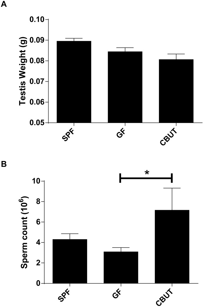
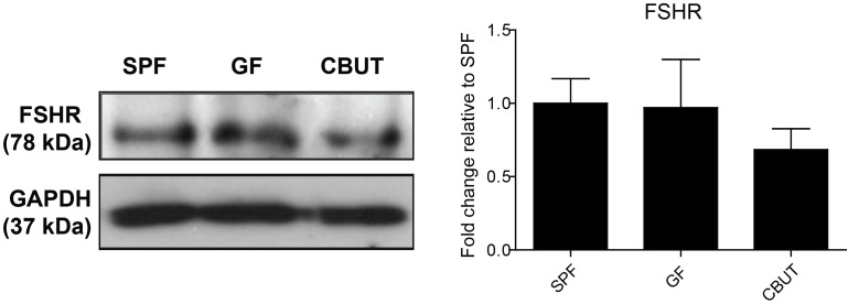
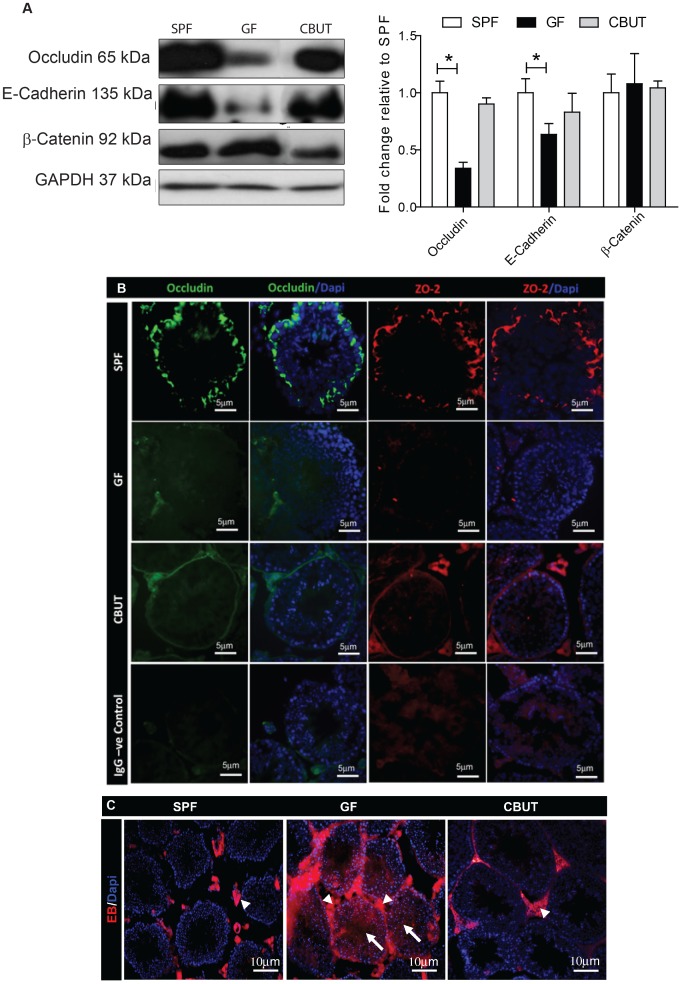
Nutrients and environmental chemicals, including endocrine disruptors, have been incriminated in the current increase in male reproductive dysfunction, but the underlying mechanisms remain unknown. The gastrointestinal tract represents the largest surface area exposed to our environment and thereby plays a key role in connection with exposure of internal organs to exogenous factors. In this context the gut microbiome (all bacteria and their metabolites) have been shown to be important contributors to body physiology including metabolism, cognitive functions and immunity. Pivotal to male reproduction is a proper development of the testis, including the formation of the blood-testis barrier (BTB) that encapsulates and protects germ cells from stress induced environmental cues, e.g. pathogenic organisms and xenobiotics. Here we used specific pathogen free (SPF) mice and germ-free (GF) mice to explore whether gut microbiota and/or their metabolites can influence testis development and regulation of BTB. Lumen formation in the seminiferous tubules, which coincides with the development of the BTB was delayed in the testes of GF mice at 16 days postpartum. In addition, perfusion experiments (Evans blue) demonstrated increased BTB permeability in these same mice. Reduced expressions of occludin, ZO-2 and E-cadherin in GF testis suggested that the microbiota modulated BTB permeability by regulation of cell-cell adhesion. Interestingly, exposure of GF mice to Clostridium Tyrobutyricum (CBUT), which secrete high levels of butyrate, restored the integrity of the BTB and normalized the levels of cell adhesion proteins. Moreover, the GF mice exhibited lower serum levels of gonadotropins (LH and FSH) than the SPF group. In addition, the intratesticular content of testosterone was lower in GF compared to SPF or CBUT animals. Thus, the gut microbiome can modulate the permeability of the BTB and might play a role in the regulation of endocrine functions of the testis.


Conflict of interest statement

Competing Interests: The authors have declared that no competing interests exist.

Figures

Figure 1
Figure 1. Testis weight (A) and sperm count (B) for SPF, GF and CBUT mice at 8 weeks of age.
The values shown are means ± SEM (n = 6) *P<0.05 calculated by the Mann-Whitney Rank Sum Test for non-parametric independent data.
Figure 2
Figure 2. Lumen formation on postnatal day 16.
(A) Representative PAS staining of seminiferous tubules from the SPF, GF and CBUT mice. (B) The percentage of open tubules and (C) tubule diameter in the seminiferous tubules of SPF, GF and CBUT mice. The values presented are means ± SEM for 9 mice from 3 different litters *P<0.05 calculated by One-way ANOVA.
Figure 3
Figure 3. Expression of Sertoli cell markers in the SPF, GF and CBUT testes.
Representative Western blots for the Follicle-stimulating hormone receptor (FSHR) and quantification of these blots relative to the levels of Glyceraldehyde 3-phosphate dehydrogenase (GAPDH) (means ± SEM, n = 3–6 *P<0.05) compared to the values for SPF mice.
Figure 4
Figure 4. Expression of tight junction proteins and changes in the permeability of the BTB of 8–10 weeks old SPF, GF and CBUT mice.
(A) Representative Western blots for occludin, E-cadherin and β-catenin, and quantification of these blots relative to the levels of GAPDH (means ± SEM., n = 3–6 *P<0.05) compared to the values for SPF mice. (B) Immunofluorescent staining for occludin (green) and ZO-2 (red) individually and merged with nuclear staining (DAPI – blue). The scale bar represents 5 µm. (C) Evans blue (EB – red) and nuclear staining (DAPI – blue) showing interstitial cells (arrowheads) and seminiferous tubules (arrows). Scale bar represents 10 µm.
Figure 5
Figure 5. Assessment of hormone levels and mRNA levels encoding steroidogenic proteins.
(A) Intratesticular levels of testosterone and serum levels of LH and FSH in 8–10 weeks old SFP, GF and CBUT mice. (a) Levels of intra-testicular testosterone per gram testis weight measured with RIA. Each bar represents the mean ± SEM (n = 6–9). The actual values were 0.315±0.131 pmol/g for SPF mice, 0.025±0.003 pmol/g for the GF group and 0.739±0.517 pmol/g for CBUT mice *P<0.05, **<0.001 as determined by the Mann-Whitney Rank Sum Test for non-parametric independent data. (b) Levels of serum LH as determined by ELISA. Each bar represents the mean ± SEM. The actual values were 179±19 ng/ml for SPF mice, 105±25 ng/ml for the GF group and 167±50 ng/ml for CBUT mice *P<0.05 as assessed with the Student t-Test for independent data. (c) Levels of serum FSH as determined by ELISA. Each bar represents mean ± SEM. The actual values were 169±18 ng/ml for SPF mice, 95±26 ng/ml for the GF group and 123±9 ng/ml for CBUT mice *P<0.05 as assessed by the Student t-Test for independent data. (B) The relative expression of mRNA species encoding steroidogenic and Leydig cell proteins normalized to the housekeeping levels of 18s rRNA and Arbpa/Rplp0 mRNA in the testes of 8–10 weeks SPF, GF and CBUT mice. Each bar represents the mean ± SEM for 6 mice from 3 different litters. cyp11a1: cytochrome P450, family 11, subfamily a, polypeptide 1, cyp19a1: cytochrome P450, family 19, subfamily a, polypeptide 1, Hsd3b1: hydroxy-delta-5-steroid dehydrogenase, 3 beta- and steroid delta-isomerase 1, Hsd17b11: hydroxysteroid (17-beta) dehydrogenase 11 and Insl3: insulin-like 3. Arbpa/Rplp0: ribosomal protein, large, P0.

References

    1. Petersen C, Soder O (2006) The sertoli cell–a hormonal target and ‘super’ nurse for germ cells that determines testicular size. Horm Res 66: 153–161. - PubMed
    1. Hess RA, Renato de Franca L (2008) Spermatogenesis and cycle of the seminiferous epithelium. Advances in experimental medicine and biology 636: 1–15. - PubMed
    1. Smith BE, Braun RE (2012) Germ cell migration across Sertoli cell tight junctions. Science 338: 798–802. - PMC - PubMed
    1. Morita K, Sasaki H, Fujimoto K, Furuse M, Tsukita S (1999) Claudin-11/OSP-based tight junctions of myelin sheaths in brain and Sertoli cells in testis. Journal of Cell Biology 145: 579–588. - PMC - PubMed
    1. Cheng CY, Mruk DD (2002) Cell junction dynamics in the testis: Sertoli-germ cell interactions and male contraceptive development. Physiological reviews 82: 825–874. - PubMed

Publication types