. 2014 Oct;10(4):1793-9.
doi: 10.3892/mmr.2014.2480. Epub 2014 Aug 11.

Rifampicin improves neuronal apoptosis in LPS-stimulated co‑cultured BV2 cells through inhibition of the TLR-4 pathway

Affiliations

Rifampicin improves neuronal apoptosis in LPS-stimulated co‑cultured BV2 cells through inhibition of the TLR-4 pathway

Wei Bi et al. Mol Med Rep. 2014 Oct.

Abstract

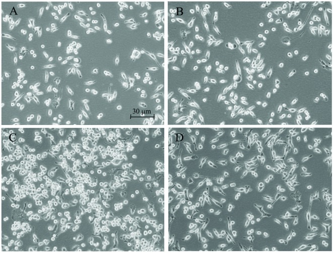
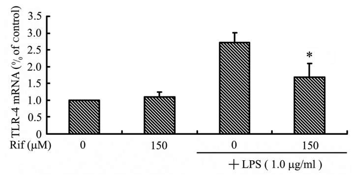
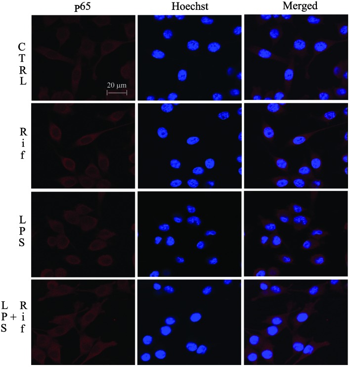
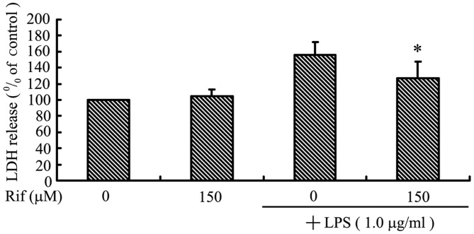
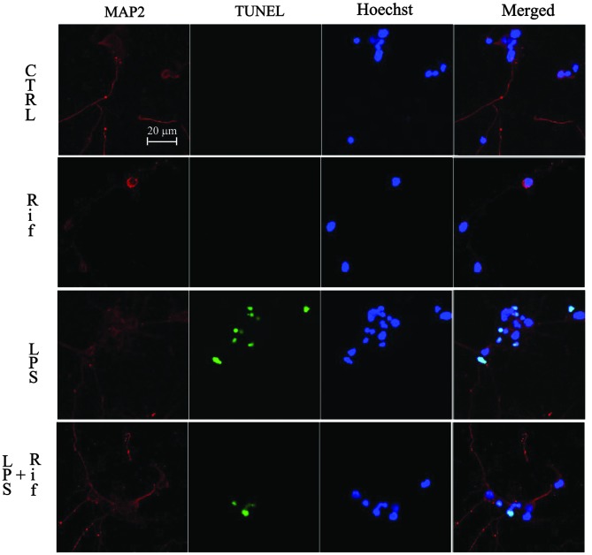
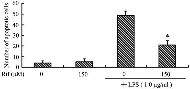
Agents inhibiting microglial activation are attracting attention as candidate drugs for neuroprotection in neurodegenerative diseases. Recently, researchers have focused on the immunosuppression induced by rifampicin. Our previous study showed that rifampicin inhibits the production of lipopolysaccharide (LPS)-induced pro-inflammatory mediators and improves neuron survival in inflammation; however, the mechanism through which rifampicin inhibits microglial inflammation and its neuroprotective effects are not completely understood. In this study, we examined the effects of rifampicin on morphological changes induced by LPS in murine microglial BV2 cells. Then we investigated, in BV2 microglia, the effects of rifampicin on two signaling pathway componentss stimulated by LPS, the Toll‑like receptor-4 (TLR-4) and the nuclear factor-κB (NF-κB). In addition, we co-cultured BV2 microglia and neurons to observe the indirect neuroprotective effects of rifampicin. Rifampicin inhibited LPS-stimulated expression of the TLR-4 gene. When neurons were co-cultured with LPS-stimulated BV2 microglia, pre-treatment with rifampicin increased neuronal viability and reduced the number of apoptotic cells. Taken together, these findings suggest that rifampicin, with its anti-inflammatory properties, may be a promising agent for the treatment of neurodegenerative diseases.


Figures

Figure 1
Figure 1
Representative images of microglia incubated for 24 h with fresh medium (control), rifampicin (150 μM) or lipopolysaccharide (LPS; 1 μg/ml). The experiments were performed on three separate microglial preparations. (A) The BV2 microglia shows the typical branching shape at the resting state. (B) Administration of rifampicin causes no obvious morphological changes compared to the non-treated group. (C) Enlargement of the microglial cell body and loss of ramifications, development of an amoeboid shape caused by LPS. (D) Treatment of cells with 150 μM rifampicin markedly improves the morphological changes caused by LPS; the microglia show a branch-like morphology.
Figure 2
Figure 2
The effects of rifampicin (Rif) on the expression of Toll-like receptor-4 (TLR-4) in lipopolysaccharide (LPS)-stimulated BV2 microglia. Reverse transcription-quantitative polymerase chain reaction analysis of TLR-4 mRNA expression. Cells were treated with 150 μM rifampicin for 2 h prior to the addition of LPS (1.0 μg/ml) for 1 h. TLR-4 mRNA levels were calculated relative to the level of the 3-phosphate dehydrogenase (GADPH) gene using standard curve analysis. Data were collected from three independent experiments, each carried out in triplicate. *P<0.05 relative to cells treated with LPS in the absence of rifampicin.
Figure 3
Figure 3
Inhibition of nuclear factor-κB (NF-κB) activation by rifampicin (Rif) in lipopolysaccharide (LPS)-stimulated BV2 microglia. Immunofluorescent staining showing the cellular distribution of the NF-κB p65 subunit (red). Cells were pre-treated with rifampicin (150 μM) for 2 h, followed by LPS treatment (1.0 μg/ml) for 2 h. Hoechst 33258 (blue) was used to visualize the nuclei. Data were collected from three independent experiments, each carried out in triplicate. Merged, double-stained slides; CTRL, control cells.
Figure 4
Figure 4
The effect of rifampicin (Rif) on cortical neuron survival in a lipopolysaccharide (LPS)-induced microglial-neuronal co-culture system. Cortical neurons were co-cultured with LPS-activated BV2 microglia with or without pre-treatment with 150 μM rifampicin for 24 h. (A) A lactate dehydrogenase (LDH) assay was used to determine the cortical neuron viability. (B) Immunofluorescent detection of apoptotic cortical neurons co-cultured with BV2 microglia. CTRL, control cells; merged, double-stained slides. (C) The number of apoptotic neurons, counted on double-stained slides from a total of 100 nuclei. Data were collected from three independent experiments, each carried out in triplicate. *p<0.05 compared to cells treated with LPS in the absence of rifampicin.
Figure 4
Figure 4
The effect of rifampicin (Rif) on cortical neuron survival in a lipopolysaccharide (LPS)-induced microglial-neuronal co-culture system. Cortical neurons were co-cultured with LPS-activated BV2 microglia with or without pre-treatment with 150 μM rifampicin for 24 h. (A) A lactate dehydrogenase (LDH) assay was used to determine the cortical neuron viability. (B) Immunofluorescent detection of apoptotic cortical neurons co-cultured with BV2 microglia. CTRL, control cells; merged, double-stained slides. (C) The number of apoptotic neurons, counted on double-stained slides from a total of 100 nuclei. Data were collected from three independent experiments, each carried out in triplicate. *p<0.05 compared to cells treated with LPS in the absence of rifampicin.

References

    1. Zhang H, Wang FW, Yao LL, Hao AJ. Microglia - friend or foe. Front Biosci (Schol Ed) 2011;3:869–883. - PubMed
    1. Smith JA, Das A, Ray SK, Banik NL. Role of pro-inflammatory cytokines released from microglia in neurodegenerative diseases. Brain Res Bull. 2012;87:10–20. - PMC - PubMed
    1. Bernardi A, Frozza RL, Meneghetti A, et al. Indomethacin-loaded lipid-core nanocapsules reduce the damage triggered by Aβ1–42 in Alzheimer’s disease models. Int J Nanomedicine. 2012;7:4927–4942. - PMC - PubMed
    1. Sung YH, Kim SC, Hong HP, et al. Treadmill exercise ameliorates dopaminergic neuronal loss through suppressing microglial activation in Parkinson’s disease mice. Life Sci. 2012;91:1309–1316. - PubMed
    1. Dibaj P, Zschüntzsch J, Steffens H, et al. Influence of methylene blue on microglia-induced inflammation and motor neuron degeneration in the SOD1(G93A) model for ALS. PLoS One. 2012;7:e43963. - PMC - PubMed

Publication types

MeSH terms