. 2014 Aug 13;9(8):e102709.
doi: 10.1371/journal.pone.0102709. eCollection 2014.

Bystander activation and anti-tumor effects of CD8+ T cells following Interleukin-2 based immunotherapy is independent of CD4+ T cell help

Affiliations

Bystander activation and anti-tumor effects of CD8+ T cells following Interleukin-2 based immunotherapy is independent of CD4+ T cell help

Arta M Monjazeb et al. PLoS One. .

Abstract

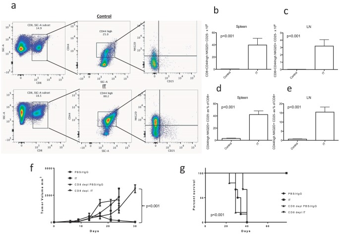
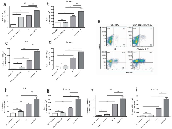
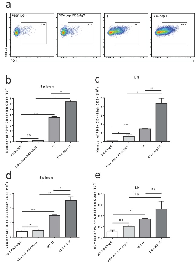
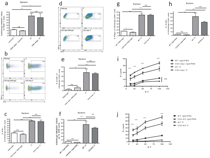
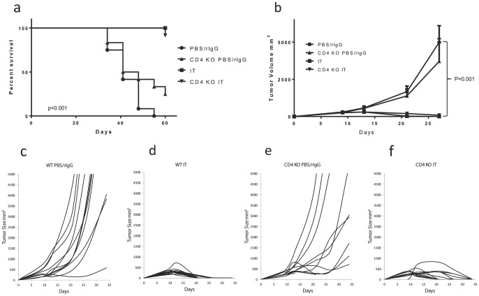
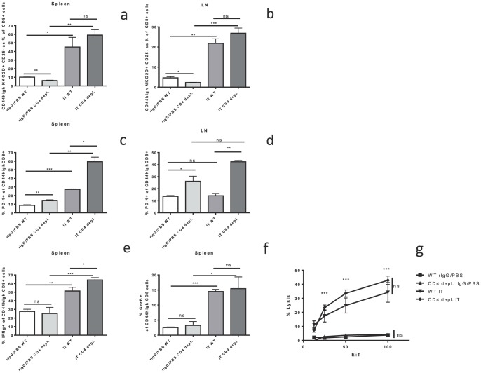
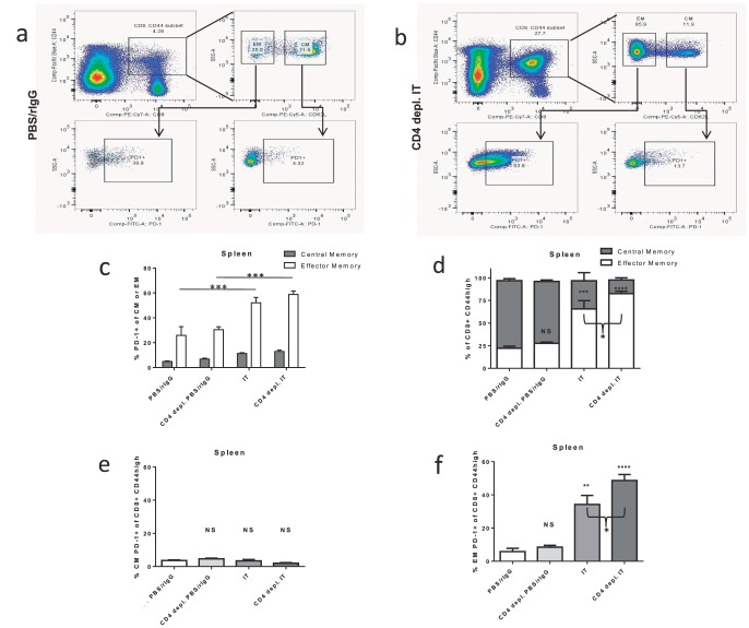
We have previously demonstrated that immunotherapy combining agonistic anti-CD40 and IL-2 (IT) results in synergistic anti-tumor effects. IT induces expansion of highly cytolytic, antigen-independent "bystander-activated" (CD8(+)CD44high) T cells displaying a CD25(-)NKG2D(+) phenotype in a cytokine dependent manner, which were responsible for the anti-tumor effects. While much attention has focused on CD4(+) T cell help for antigen-specific CD8(+) T cell expansion, little is known regarding the role of CD4(+) T cells in antigen-nonspecific bystander-memory CD8(+) T cell expansion. Utilizing CD4 deficient mouse models, we observed a significant expansion of bystander-memory T cells following IT which was similar to the non-CD4 depleted mice. Expanded bystander-memory CD8(+) T cells upregulated PD-1 in the absence of CD4(+) T cells which has been published as a hallmark of exhaustion and dysfunction in helpless CD8(+) T cells. Interestingly, compared to CD8(+) T cells from CD4 replete hosts, these bystander expanded cells displayed comparable (or enhanced) cytokine production, lytic ability, and in vivo anti-tumor effects suggesting no functional impairment or exhaustion and were enriched in an effector phenotype. There was no acceleration of the post-IT contraction phase of the bystander memory CD8(+) response in CD4-depleted mice. The response was independent of IL-21 signaling. These results suggest that, in contrast to antigen-specific CD8(+) T cell expansion, CD4(+) T cell help is not necessary for expansion and activation of antigen-nonspecific bystander-memory CD8(+) T cells following IT, but may play a role in regulating conversion of these cells from a central memory to effector phenotype. Additionally, the expression of PD-1 in this model appears to be a marker of effector function and not exhaustion.


Conflict of interest statement

Competing Interests: The authors have declared that no competing interests exist.

Figures

Figure 1
Figure 1. CD40/IL-2 Immunotherapy induces massive expansion of bystander memory CD8+ cells and anti-tumor effects are CD8 dependent.
Three C57BL/6 mice per group were treated with IT or PBS/rIgG (control) and effects on CD8+ T cell expansion were quantified by flow cytometric analysis 11 days after the initiation of therapy. For in vivo tumor studies one million 3LL cells were administered by s.c. injection into the flank of C57BL/6 mice seven days prior to initiation of therapy. Six to eight 3LL bearing mice were treated with IT and/or CD8+ T cell depletion to examine CD8+ dependence of anti-tumor effects. (a) Gating strategy for bystander memory CD8+ CD44high NKG2D+ CD25− cells. (b–e) Expansion of bystander memory CD8+ T cells in the spleen and lymph nodes of IT or vehicle treated mice expressed as total numbers (b,c) or as a percentage of total CD8+ T cells (d,e). Effects of IT and/or CD8 depletion on tumor growth (f) and survival (g).
Figure 2
Figure 2. IT induced expansion of memory CD8+ T cells in CD4+ T cell deficient models.
Control or CD4 deficient (depleted or knockout) C57BL/6 mice were treated with IT or PBS/rIgG (control) and effects on CD8+ T cell expansion were quantified by flow cytometric analysis 11 days after the initiation of IT. CD8+ (a,b) and memory CD8+ (c,d) T cell numbers in the LNs (a,c) and spleens (b,d) of control or CD4+ T cell depleted mice treated with vehicle or IT. (e) BrdU incorporation in CD8+ T cells from spleens of control or CD4+ T cell depleted mice treated with vehicle or IT. CD8+ (f,g) and memory CD8+ (h,i) T cell numbers in the LNs (f,h) and spleens (g,i) of wild-type or CD4 knockout mice treated with vehicle or IT. Results are representative of two (CD4 knockout) or three (CD4 depletion) independent experiments with a minimum of three mice per group. (*P<.05, **P<.01, ***P<.001).
Figure 3
Figure 3. Increased number of PD-1+ memory CD8+ T cells after IT in CD4+ T cell deficient mice.
Control or CD4 deficient (depleted or knockout) C57BL/6 mice were treated with IT or PBS/rIgG (control) and PD-1 expression on memory T-cells was quantified by flow cytometric analysis 11 days after the initiation of IT. (a) Representative dot plots for PD-1+ gating on CD8+ CD44high cells in the spleens of CD4+ depletion model mice. Number of PD-1+ memory (CD44high) CD8+ T cells in spleens (b,d) and LNs+ (c,e) of IT or vehicle treated mice in CD4+ depletion (b,c) or CD4 knockout (d,e) models. Results are representative of two (CD4 knockout) or three (CD4 depletion) independent experiments with a minimum of three mice per group. (*P<.05, **P<.01, ***P<.001).
Figure 4
Figure 4. Memory CD8+ T cell function after IT in CD4+ T cell deficient models.
Control or CD4 deficient (depleted or knockout) C57BL/6 mice were treated with IT or PBS/rIgG (control) and assessed for function of memory CD8+ T cells 11 days after the initiation of IT. NKG2D and granzyme B expression were quantified by flow cytometric analysis. Interferon gamma production was quantified by flow cytometric analysis after in vitro restimulation of splenocytes with PMA/Ionomycin (0.16/1.6 ug/ml) for one hour followed by incubation with golgi stop (0.7 ug/ml) for three hours. CD8+ T cell killing function was assayed by scintillation counting using an in vitro redirected lysis assay with 51Cr labeled P815 target cells incubated for 30 minutes with 10 ug/mL anti-CD3e. (a,f) NKG2D expression on memory CD8+ T cells in CD4 depletion (a) and knockout (f) models. Representative dot plots for NKG2D+ CD25− gating are presented in Figure 1. (b) Representative dot plots for IFNγ+ gating on CD8+ CD44high cells in the spleens of CD4+ depletion model mice. (c,g) Interferon gamma production by memory CD8+ T cells in CD4 depletion (c) and knockout (g) models. (d) Representative dot plots for Granzyme B+ gating on CD8+ CD44high cells in the spleens of CD4+ depletion model mice. (e,h) Granzyme B expression by memory CD8+ T cells in CD4 depletion (e) and knockout (h) models. Killing function of splenocytes from CD4 depleted (i) or CD knockout (j) mice expressed as percentage of maximal lysis. Results are representative of two (CD4 knockout) or three (CD4 depletion) independent experiments with a minimum of three mice per group. (*P<.05, **P<.01, ***P<.001).
Figure 5
Figure 5. Anti-tumor effects of IT in CD4 knockout mice.
3LL tumor bearing WT or CD4 knockout (B6.129S2-CD4tm1Mak/J) mice were treated with IT or PBS/rIgG (control) and survival and tumor growth were measured. For in vivo tumor studies one million 3LL cells were administered by s.c. injection into the flank of C57BL/6 mice seven days prior to initiation of therapy. (a) Survival. (b) Mean tumor volume with SEM. (c–f) Growth plots of individual tumors in each group. N = 12 mice per group. (*P<.05, **P<.01, ***P<.001).
Figure 6
Figure 6. CD4 independence of IT induced bystander activated memory CD8+ T cells persists at longer time points.
Control or long term CD4+ depleted C57BL/6 mice were treated with IT or PBS/rIgG (control) and harvested at Day 15 after the initiation of therapy. Bystander memory CD8+ T cells, PD-1 expression, interferon gamma production, and granzyme B production were quantified by flow cytometric analysis. Cytotoxic effector function was assayed by a redirected lysis assay. The percentage of CD8+ T cells with the CD44high NKG2D+ CD25− bystander memory phenotype in the spleen (a) and LNs (b). PD-1 expression on the memory CD8+ T cells in the spleen (c) and LNs (d). Interferon gamma production (e) and granzyme B expression (f) in splenic memory CD8+ T cells. (g) Killing function of splenocytes from long term CD4 depleted mice expressed as percentage of maximal lysis. N = 3 mice per group (*P<.05, **P<.01, ***P<.001).
Figure 7
Figure 7. PD-1 expression on central and effector memory CD8+ T cells after IT in CD4+ T cell depleted mice.
Control or CD4+ depleted C57BL/6 mice were treated with IT or PBS/rIgG (control). CD62L and PD-1 expression on memory T-cells was quantified by flow cytometric analysis 11 days after the initiation of IT. (a,b) Examples of the gating strategy for PD-1 expression on the CM and EM components of the memory CD8+ T cell compartment. The majority of cells in the memory compartment are CM in untreated mice (a) and EM in IT treated mice (b). (c) PD-1 expression on CM and EM cells. (d) The composition of the memory CD8+ T cell compartment in control or CD4 depleted mice treated with IT or PBS/rIgG. PD-1+ CM cells (e) and PD-1+ EM cells (f) as a percentage of the total memory CD8+ T cell compartment. Results are representative of two to three independent experiments with 3 mice per group (*P<.05, **P<.01, ***P<.001, ****P<.0001).

References

    1. Murali-Krishna K, Altman JD, Suresh M, Sourdive DJ, Zajac AJ, et al. (1998) Counting antigen-specific CD8 T cells: a reevaluation of bystander activation during viral infection. Immunity 8: 177–187. - PubMed
    1. Tough DF, Borrow P, Sprent J (1996) Induction of bystander T cell proliferation by viruses and type I interferon in vivo. Science 272: 1947–1950. - PubMed
    1. Tietze JK, Wilkins DE, Sckisel GD, Bouchlaka MN, Alderson KL, et al. (2012) Delineation of antigen-specific and antigen-nonspecific CD8(+) memory T-cell responses after cytokine-based cancer immunotherapy. Blood 119: 3073–3083. - PMC - PubMed
    1. Belz GT, Doherty PC (2001) Virus-specific and bystander CD8+ T-cell proliferation in the acute and persistent phases of a gammaherpesvirus infection. J Virol 75: 4435–4438. - PMC - PubMed
    1. Dhanji S, Teh SJ, Oble D, Priatel JJ, Teh HS (2004) Self-reactive memory-phenotype CD8 T cells exhibit both MHC-restricted and non-MHC-restricted cytotoxicity: a role for the T-cell receptor and natural killer cell receptors. Blood 104: 2116–2123. - PubMed

Publication types

MeSH terms