. 2014 Aug 1:11:23.
doi: 10.1186/1742-6405-11-23. eCollection 2014.

Differential glycosylation of envelope gp120 is associated with differential recognition of HIV-1 by virus-specific antibodies and cell infection

Affiliations

Differential glycosylation of envelope gp120 is associated with differential recognition of HIV-1 by virus-specific antibodies and cell infection

Milan Raska et al. AIDS Res Ther. .

Abstract

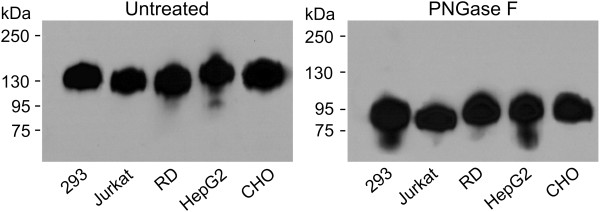
Background: HIV-1 entry into host cells is mediated by interactions between the virus envelope glycoprotein (gp120/gp41) and host-cell receptors. N-glycans represent approximately 50% of the molecular mass of gp120 and serve as potential antigenic determinants and/or as a shield against immune recognition. We previously reported that N-glycosylation of recombinant gp120 varied, depending on the producer cells, and the glycosylation variability affected gp120 recognition by serum antibodies from persons infected with HIV-1 subtype B. However, the impact of gp120 differential glycosylation on recognition by broadly neutralizing monoclonal antibodies or by polyclonal antibodies of individuals infected with other HIV-1 subtypes is unknown.

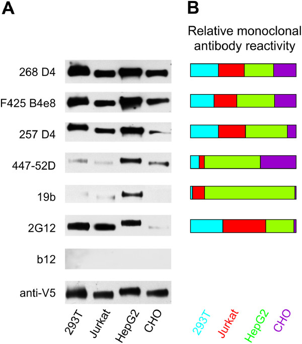
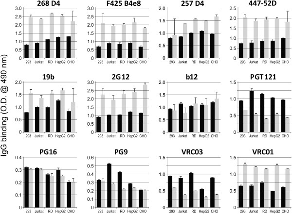
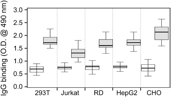
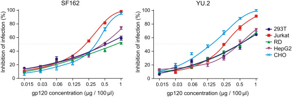
Methods: Recombinant multimerizing gp120 antigens were expressed in different cells, HEK 293T, T-cell, rhabdomyosarcoma, hepatocellular carcinoma, and Chinese hamster ovary cell lines. Binding of broadly neutralizing monoclonal antibodies and polyclonal antibodies from sera of subtype A/C HIV-1-infected subjects with individual gp120 glycoforms was assessed by ELISA. In addition, immunodetection was performed using Western and dot blot assays. Recombinant gp120 glycoforms were tested for inhibition of infection of reporter cells by SF162 and YU.2 Env-pseudotyped R5 viruses.

Results: We demonstrated, using ELISA, that gp120 glycans sterically adjacent to the V3 loop only moderately contribute to differential recognition of a short apex motif GPGRA and GPGR by monoclonal antibodies F425 B4e8 and 447-52D, respectively. The binding of antibodies recognizing longer peptide motifs overlapping with GPGR epitope (268 D4, 257 D4, 19b) was significantly altered. Recognition of gp120 glycoforms by monoclonal antibodies specific for other than V3-loop epitopes was significantly affected by cell types used for gp120 expression. These epitopes included CD4-binding site (VRC03, VRC01, b12), discontinuous epitope involving V1/V2 loop with the associated glycans (PG9, PG16), and an epitope including V3-base-, N332 oligomannose-, and surrounding glycans-containing epitope (PGT 121). Moreover, the different gp120 glycoforms variably inhibited HIV-1 infection of reporter cells.

Conclusion: Our data support the hypothesis that the glycosylation machinery of different cells shapes gp120 glycosylation and, consequently, impacts envelope recognition by specific antibodies as well as the interaction of HIV-1 gp120 with cellular receptors. These findings underscore the importance of selection of appropriately glycosylated HIV-1 envelope as a vaccine antigen.

Keywords: Deglycosylation resistance; Glycan-specific antibody; Neutralization inhibition; gp120 glycosylation.


Figures

Figure 1
Figure 1
Reactivities of Env-specific monoclonal antibodies with gp120 produced in different cell lines determined by Western blot. (A) Antibody binding to gp120 glycoforms produced in HEK 293T, Jurkat, HepG2, and CHO cell lines was analyzed by Western blot after SDS-PAGE separation under reducing conditions. Western blots were developed with monoclonal antibodies 268 D4, F425 B4e8, 257 D4, 447-52D, 19b, 2G12, and b12. Anti-V5-tag antibody was used to assess the amount of loaded gp120. (B) Densities of individual gp120 glycoform bands obtained from two Western blots after developing with individual monoclonal antibodies were analyzed by ImageJ 1.41a software. Densitometric values were normalized to loaded amount of individual gp120 glycoforms (V5-tag-positive bands). Mean values from two Western blots were calculated and expressed as relative reactivity of individual monoclonal antibodies with each gp120 glycoform.
Figure 2
Figure 2
Comparison of reactivities of selected HIV-1 gp120-specific monoclonal antibodies with native and deglycosylated recombinant HIV-1 gp120 analyzed by ELISA. ELISA plates were coated with anti-penta-His antibody followed by capture of equal amounts of gp120 produced in HEK 293T (abbreviated as 293), Jurkat, RD, HepG2, or CHO cells (0.05 μg gp120 per well). gp120 preparations were in native form (black columns) or deglycosylated with PNGase F (grey columns). Monoclonal antibodies 268 D4, F425 B4e8, 257 D4, 447-52D, 19b, 2G12, b12, PGT121, PG16, PG9, VRC03, and VRC01 were added and the bound IgG antibodies were detected with HRP-conjugated goat anti-human IgG. Optimal dilutions for each antibody were determined in preliminary experiments using gp120 produced in HEK 293T cells and it is specified in the Table 1. Values correspond to mean absorbance ± SD. Averaged results from three experiments with independently produced recombinant gp120 glycoforms are shown.
Figure 3
Figure 3
Mobility shift of gp120 after deglycosylation. gp120 glycoproteins produced in HEK 293T, Jurkat, RD, HepG2, and CHO cells were deglycosylated by PNGase F under native conditions, separated by SDS-PAGE under reducing conditions, and detected with anti-V5-tag monoclonal antibody. Change in migration of untreated (left panel) vs. PNGase F-treated (right panel) gp120 preparations was approximately from 130 kDa to 85 KDa. Results from one of two experiments are shown.
Figure 4
Figure 4
Reactivities of serum IgG from HIV-1-infected subjects with HIV-1 gp120 produced in different cell lines analyzed by Western blot (A, C) and dot-blot (B, D). HIV-1 Env gp120 glycoforms produced in HEK 293T, Jurkat, RD, HepG2 and CHO cells were (A) separated by SDS-PAGE under reducing conditions and blotted onto a PVDF membrane or (B) dot blotted onto a PVDF membrane and both were developed with sera (0.2 μg/ml gp120-specific IgG) from HIV-1-infected subjects (P1-9; HIV-1+ sera) or sera (1:200 dilution) from healthy sero-negative subjects (C1-3; Control sera). Results from one of two experiments are shown. (C, D) Densitometric analysis performed with the ImageJ program shows mean reactivity of each serum IgG with individual gp120 variants.
Figure 5
Figure 5
Reactivities of serum IgG from HIV-1-infected subjects with native and deglycosylated HIV-1 gp120. ELISA plates were coated with anti-penta-His capture antibody followed by binding of equal amounts of gp120 produced in HEK 293T, Jurkat, RD, HepG2, or CHO cells (0.05 μg per well). Captured gp120 proteins were native (empty boxes) or deglycosylated (gray-filled boxes) using PNGase F under native conditions. Sera from nine HIV-1-infected subjects were added to the ELISA plates at predetermined dilutions (see Methods). Bound IgG antibodies were detected with HRP-conjugated goat anti-human IgG. Data from three independent experiments are shown. Thick line represents median values and boxes the values between 25th and 75th percentiles. The upper and lower whiskers limit 95% of measured values.
Figure 6
Figure 6
Inhibition of SF162 and YU.2 HIV-1 infectivity by differentially glycosylated recombinant gp120. SF162 or YU.2 pseudoviruses (200 TCID50) together with serially diluted gp120 preparations (1 to 0.015 μg/well) were added to TZM-bl reporter cells, and 48 h later luciferase activity was measured in the cell lysates. Results are expressed as % inhibition of the infection of TZM-bl reporter cells, with pseudovirus alone considered 100% infection. Data from three independent experiments are shown.
Figure 7
Figure 7
Inhibition of SF162 and YU.2 HIV-1 infectivity by differentially glycosylated recombinant gp120 preincubated with TZM-bl indicator cells. SF162 and YU.2 pseudoviruses were added to TZM-bl reporter cells pre-incubated with serially diluted gp120 preparations (1 to 0.015 μg/well). The inhibition of the infectivity was determined and expressed as in Figure 6. Data from three independent experiments are shown.

References

    1. Kwong PD, Wyatt R, Robinson J, Sweet RW, Sodroski J, Hendrickson WA. Structure of an HIV gp120 envelope glycoprotein in complex with the CD4 receptor and a neutralizing human antibody. Nature. 1998;393:648–659. - PMC - PubMed
    1. Wyatt R, Kwong PD, Desjardins E, Sweet RW, Robinson J, Hendrickson WA, Sodroski JG. The antigenic structure of the HIV gp120 envelope glycoprotein. Nature. 1998;393:705–711. - PubMed
    1. Pantophlet R, Burton DR. gp120: target for neutralizing HIV-1 antibodies. Annu Rev Immunol. 2006;24:739–769. - PubMed
    1. Wei X, Decker JM, Wang S, Hui H, Kappes JC, Wu X, Salazar-Gonzalez JF, Salazar MG, Kilby JM, Saag MS, Komarova NL, Nowak MA, Hahn BH, Kwong PD, Shaw GM. Antibody neutralization and escape by HIV-1. Nature. 2003;422:307–312. - PubMed
    1. Arthos J, Cicala C, Martinelli E, Macleod K, Van Ryk D, Wei D, Xiao Z, Veenstra TD, Conrad TP, Lempicki RA, McLaughlin S, Pascuccio M, Gopaul R, McNally J, Cruz CC, Censoplano N, Chung E, Reitano KN, Kottilil S, Goode DJ, Fauci AS. HIV-1 envelope protein binds to and signals through integrin alpha4beta7, the gut mucosal homing receptor for peripheral T cells. Nat Immunol. 2008;9:301–309. - PubMed