

Accuracy of fine needle aspiration biopsy processed by cytologic smear and cell block techniques for the diagnosis of lacrimal gland tumors: a study of 48 cases
- PMID: 25120744
- PMCID: PMC4128979
Accuracy of fine needle aspiration biopsy processed by cytologic smear and cell block techniques for the diagnosis of lacrimal gland tumors: a study of 48 cases
Abstract
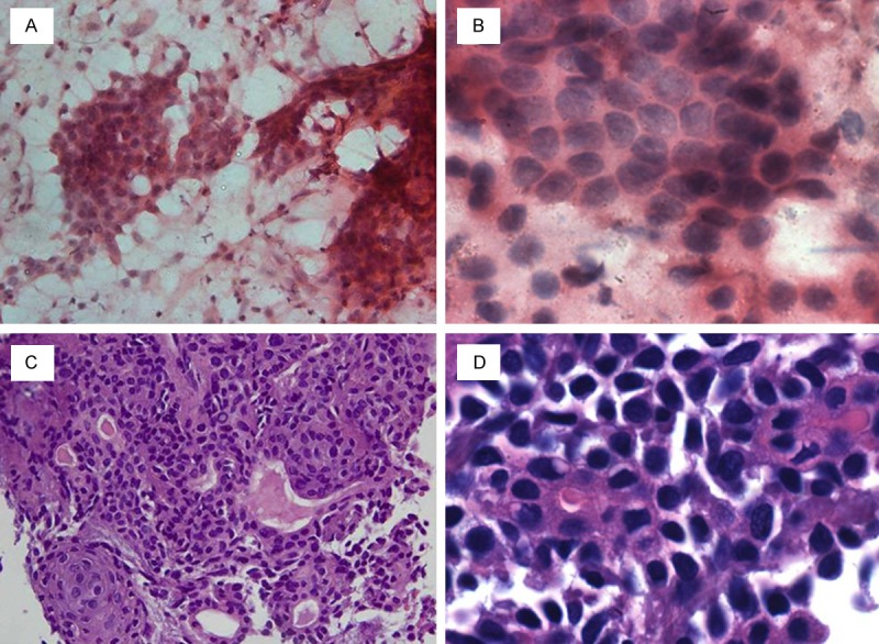
Objective: To study the accuracy of fine needle aspiration biopsy (FNAB) processed by smear cytology and cell block (CB) techniques for the diagnosis of lacrimal gland tumors (LGTs).
Study design: In a prospective study, we enrolled 48 consecutive patients with LGTs. Immediately after excision of LGTs, the tissues were underwent FNAB with 23-gauge needles. The FNAB samples were processed to produce cytologic smears and CB from which slides were cut for immunohistochemical staining. The remainders were submitted for routine histopathologic processing. The diagnostic value of FNAB was assessed by comparing the FNAB diagnoses to those made by routine histopathology.
Results: Cytopathologic evaluations based on smear cytology and CB with sections stained immunohistochemically can distinguish non-epithelial lesions from epithelial ones in all cases. The diagnostic sensitivities, specificities, and accuracies for distinguishing benign from malignant lesions were: cytologic smears--76%, 68%, and 71%, respectively; CB with immunohistochemical staining--88%, 87%, and 88%, respectively. The accuracy of the tissue diagnosis compared to routine histopathology was less for cytologic smears (58%) than for CB with immunohistochemistry (81%; P < 0.05).
Conclusions: FNAB of LGT processed using a CB technique capable of producing immunohistochemically stained slides results in a greater percentage of accurate tissue diagnoses than do cytologic smears, when compared to routine histopathology.
Keywords: Lacrimal gland lesions; cell blocks; cytopathology; fine needle aspiration; immnohistichemistry.
Figures


Similar articles
-
Cell Block-Based Two-Dimensional and Immunocytochemical Analyses Could Reduce Atypical/Indeterminate Case Frequency in Breast Fine-Needle Aspiration Cytology: A Retrospective Analysis.Acta Cytol. 2023;67(6):583-592. doi: 10.1159/000534517. Epub 2023 Oct 11. Acta Cytol. 2023. PMID: 37820610
-
The role of fine needle aspiration biopsy in deep lobe parotid tumors: Comparison of preoperative cytology and postoperative histopathologic results.Am J Otolaryngol. 2021 Jan-Feb;42(1):102590. doi: 10.1016/j.amjoto.2020.102590. Epub 2020 Jun 10. Am J Otolaryngol. 2021. PMID: 33045535
-
Sonographically guided fine-needle aspiration biopsy of major salivary gland masses: a review of 245 cases.AJR Am J Roentgenol. 2011 May;196(5):1160-3. doi: 10.2214/AJR.10.4256. AJR Am J Roentgenol. 2011. PMID: 21512086
-
Systematic Review and Meta-Analysis of the Diagnostic Accuracy of the International Academy of Cytology Yokohama System for Reporting Breast Fine-Needle Aspiration Biopsy in Diagnosing Breast Cancer.Acta Cytol. 2023;67(1):1-16. doi: 10.1159/000527346. Epub 2022 Nov 22. Acta Cytol. 2023. PMID: 36412573
-
Breast fine needle aspiration biopsy cytology: the potential impact of the International Academy of Cytology Yokohama System for Reporting Breast Fine Needle Aspiration Biopsy Cytopathology and the use of rapid on-site evaluation.J Am Soc Cytopathol. 2020 Mar-Apr;9(2):103-111. doi: 10.1016/j.jasc.2019.10.004. Epub 2020 Jan 23. J Am Soc Cytopathol. 2020. PMID: 32044283 Review.
References
-
- Shields JA, Shields CL, Scartozzi R. Survey of 1264 patients with orbital tumors and simulating lesions: The 2002 Montgomery Lecture, part 1. Ophthalmology. 2004;111:997–1008. - PubMed
-
- Prabhakaran VC, Cannon PS, McNab A, Davis G, O’Donnell B, Dolman PJ, Ghabrial R, Selva D. Lesions mimicking lacrimal gland pleomorphic adenoma. Br J Ophthalmol. 2010;94:1509–1512. - PubMed
-
- von Holstein SL, Coupland SE, Briscoe D, Le Tourneau C, Heegaard S. Epithelial tumours of the lacrimal gland: a clinical, histopathological, surgical and oncological survey. Acta Ophthalmol. 2013;91:195–206. - PubMed
MeSH terms
LinkOut - more resources
Full Text Sources
Medical