

Propranolol enhanced adipogenesis instead of induction of apoptosis of hemangiomas stem cells
- PMID: 25120757
- PMCID: PMC4128992
Propranolol enhanced adipogenesis instead of induction of apoptosis of hemangiomas stem cells
Abstract
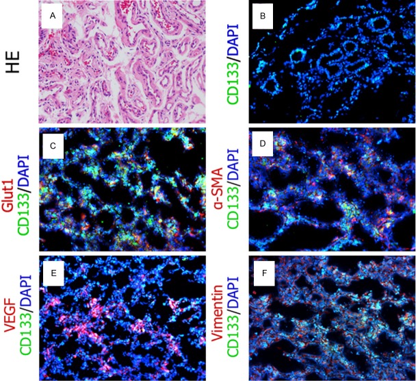
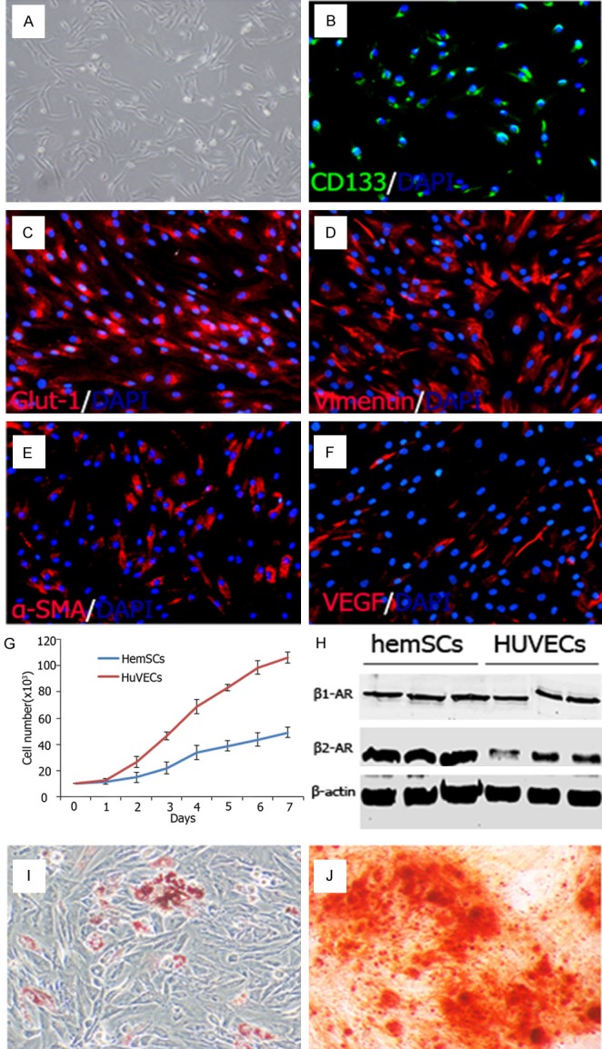
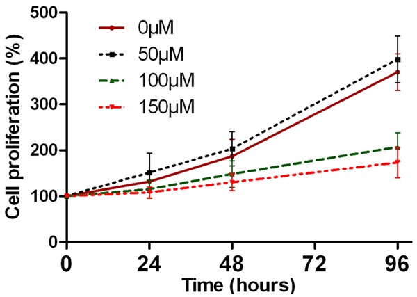
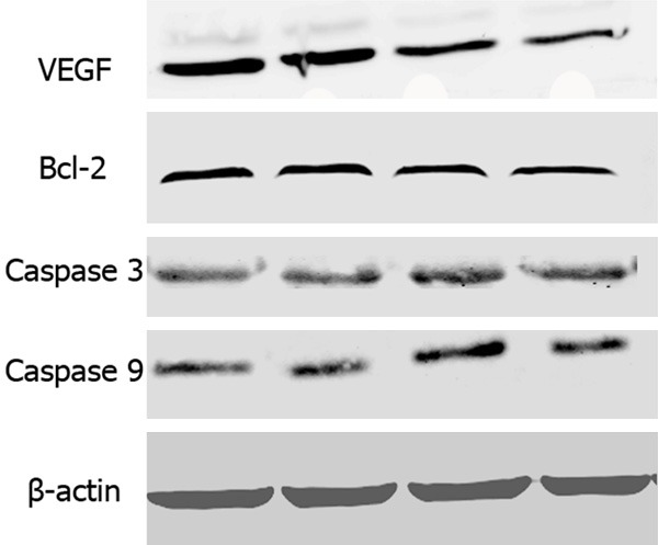
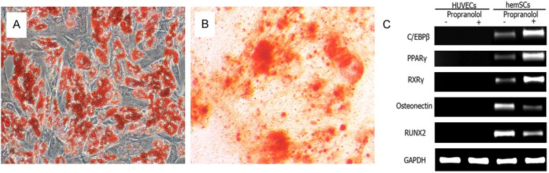
Propranolol has been widely used in treating infantile hemangiomas (IHs). But recurrence of IHs was found in some cases on cessation of propranolol treatment. The other is that Chinese individuals reacted to propranolol differently from American Whites. Whether the difference of sensitivity is due to the β adrenoceptor (β-AR) expression pattern of hemangioma initiating cells remains unclear. In the present study, we isolated hemangioma-derived stem cells (hemSCs) from proliferative IHs and analyzed the biological characteristics and β-AR expression pattern of hemSCs by immunostaining, Western blotting and multilineage differentiation assay as well. We also tested the effects of propranolol on hemSCs by evaluating VEGF expression, proliferation and apoptosis related parameters. Our results indicated that CD133(+) hemSCs located pre-vascular in proiferative IH tissues. Both β1 and β2-AR were expressed, while β2-AR was dominant on hemSCs. Propranolol at 100-150 μM inhibited proliferation of hemSCs, not did 50 μM. Propranolol down-regulated VEGF expression of hemSCs, instead of inducing apoptosis. The adipogenic potential was enhanced by propranolol. Therefore, our current results suggested propranolol could not induce apoptosis of hemSCs, but played a curative role though suppressing VEGF synthesis and enhancement of adipogenesis of hemSCs. Our results might partially provide the insight of mechanism of relapse in some cases on cessation of propranolol treatment.
Keywords: Propranolol; hemangioma-stem cells; relapse; β adrenoceptor.
Figures
Similar articles
-
Propranolol inhibits angiogenesis via down-regulating the expression of vascular endothelial growth factor in hemangioma derived stem cell.Int J Clin Exp Pathol. 2013 Dec 15;7(1):48-55. eCollection 2014. Int J Clin Exp Pathol. 2013. PMID: 24427325 Free PMC article.
-
Propranolol Targets Hemangioma Stem Cells via cAMP and Mitogen-Activated Protein Kinase Regulation.Stem Cells Transl Med. 2016 Jan;5(1):45-55. doi: 10.5966/sctm.2015-0076. Epub 2015 Nov 16. Stem Cells Transl Med. 2016. PMID: 26574555 Free PMC article.
-
Propranolol Accelerats Hemangioma Stem Cell Transformation Into Adipocyte.Ann Plast Surg. 2019 Nov;83(5):e5-e13. doi: 10.1097/SAP.0000000000002104. Ann Plast Surg. 2019. PMID: 31609806
-
Infantile hemangiomas, retinopathy of prematurity and cancer: a common pathogenetic role of the β-adrenergic system.Med Res Rev. 2015 May;35(3):619-52. doi: 10.1002/med.21336. Epub 2014 Dec 19. Med Res Rev. 2015. PMID: 25523517 Review.
-
Propranolol treatment in life-threatening airway hemangiomas: a case series and review of literature.Int J Pediatr Otorhinolaryngol. 2013 Nov;77(11):1791-800. doi: 10.1016/j.ijporl.2013.08.011. Epub 2013 Aug 22. Int J Pediatr Otorhinolaryngol. 2013. PMID: 24074695 Review.
Cited by
-
CD44-positive cancer stem cells from oral squamous cell carcinoma exhibit reduced proliferation and stemness gene expression upon adipogenic induction.Med Oncol. 2022 Jan 4;39(2):23. doi: 10.1007/s12032-021-01617-4. Med Oncol. 2022. PMID: 34982245
-
β-Adrenoceptors in Cancer: Old Players and New Perspectives.Handb Exp Pharmacol. 2024;285:665-688. doi: 10.1007/164_2023_701. Handb Exp Pharmacol. 2024. PMID: 37982890 Review.
-
Modulation of LIN28B/Let-7 Signaling by Propranolol Contributes to Infantile Hemangioma Involution.Arterioscler Thromb Vasc Biol. 2018 Jun;38(6):1321-1332. doi: 10.1161/ATVBAHA.118.310908. Epub 2018 May 3. Arterioscler Thromb Vasc Biol. 2018. PMID: 29724816 Free PMC article.
-
Propranolol inhibits stemness of hemangioma through Jagged1.Ann Transl Med. 2021 Nov;9(22):1682. doi: 10.21037/atm-21-5563. Ann Transl Med. 2021. PMID: 34988191 Free PMC article.
-
The cytotoxic and apoptotic effects of beta-blockers with different selectivity on cancerous and healthy lung cell lines.Mol Biol Rep. 2021 May;48(5):4009-4019. doi: 10.1007/s11033-021-06409-7. Epub 2021 Jun 16. Mol Biol Rep. 2021. PMID: 34136985
References
-
- Itinteang T, Brasch HD, Tan ST, Day DJ. Expression of components of the renin-angiotensin system in proliferating infantile haemangioma may account for the propranolol-induced accelerated involution. J Plast Reconstr Aesthet Surg. 2011;64:759–765. - PubMed
-
- Haggstrom AN, Drolet BA, Baselga E, Chamlin SL, Garzon MC, Horii KA, Lucky AW, Mancini AJ, Metry DW, Newell B, Nopper AJ, Frieden IJ. Prospective study of infantile hemangiomas: clinical characteristics predicting complications and treatment. Pediatrics. 2006;118:882–887. - PubMed
-
- Leaute-Labreze C, Dumas de la Roque E, Hubiche T, Boralevi F, Thambo JB, Taieb A. Propranolol for severe hemangiomas of infancy. N Engl J Med. 2008;358:2649–2651. - PubMed
-
- Ma X, Zhao T, Xiao Y, Yu J, Chen H, Huang Y, Liu J, Lin J, Ouyang T. Preliminary experience on treatment of infantile hemangioma with low-dose propranolol in China. Eur J Pediatr. 2013;172:653–659. - PubMed
-
- Ji Y, Li K, Xiao X, Zheng S, Xu T, Chen S. Effects of propranolol on the proliferation and apoptosis of hemangioma-derived endothelial cells. J Pediatr Surg. 2012;47:2216–2223. - PubMed
Publication types
MeSH terms
Substances
LinkOut - more resources
Full Text Sources
Other Literature Sources
Medical
Research Materials