Review
. 2014 Aug 10:11:18.
doi: 10.1186/2045-8118-11-18. eCollection 2014.

Vascular disruption and blood-brain barrier dysfunction in intracerebral hemorrhage

Affiliations
Review

Vascular disruption and blood-brain barrier dysfunction in intracerebral hemorrhage

Richard F Keep et al. Fluids Barriers CNS. .

Abstract

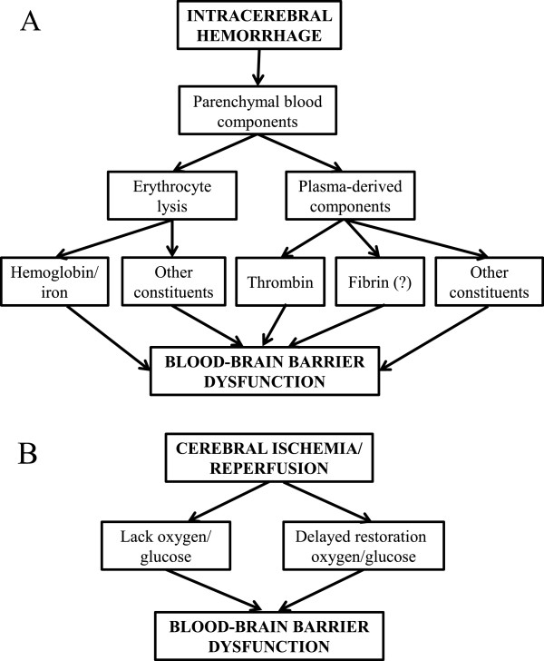
This article reviews current knowledge of the mechanisms underlying the initial hemorrhage and secondary blood-brain barrier (BBB) dysfunction in primary spontaneous intracerebral hemorrhage (ICH) in adults. Multiple etiologies are associated with ICH, for example, hypertension, Alzheimer's disease, vascular malformations and coagulopathies (genetic or drug-induced). After the initial bleed, there can be continued bleeding over the first 24 hours, so-called hematoma expansion, which is associated with adverse outcomes. A number of clinical trials are focused on trying to limit such expansion. Significant progress has been made on the causes of BBB dysfunction after ICH at the molecular and cell signaling level. Blood components (e.g. thrombin, hemoglobin, iron) and the inflammatory response to those components play a large role in ICH-induced BBB dysfunction. There are current clinical trials of minimally invasive hematoma removal and iron chelation which may limit such dysfunction. Understanding the mechanisms underlying the initial hemorrhage and secondary BBB dysfunction in ICH is vital for developing methods to prevent and treat this devastating form of stroke.

Keywords: Blood–brain barrier; Endothelium; Hematoma expansion; Hemoglobin; Intracerebral hemorrhage; Iron; Thrombin; Tight junction.


Figures

Figure 1
Figure 1
Photomicrographs showing both sides of serial coronal sections (thickness approximately 5 mm) containing hematoma (#) and edematous white matter (*) 24 hours after blood infusion in a pig brain frozen in situ. Perihematomal edema is present as blue-staining translucent regions in white matter adjacent to the hematoma. Evans blue staining is observed throughout the ipsilateral white matter and is indicative of increased BBB permeability and vasogenic edema development. Figure/legend reprinted with permission from Journal of Neurosurgery[11].
Figure 2
Figure 2
Flair magnetic resonance imaging in a patient at day 1 after ICH. Note the marked edema (#) surrounding the hematoma (*).
Figure 3
Figure 3
Hematoxylin & eosin staining of a section from the brain of a patient with cerebral cavernous malformation (type CCM3). Note the multiple blood vessels with little or no intervening parenchymal tissue. Scale bar = 200 μm.
Figure 4
Figure 4
The underlying cause of secondary BBB dysfunction appears to differ in ICH (A) from cerebral ischemia (B). In ICH, evidence indicates a major role for the presence of blood components in brain parenchyma activating a number of pathways (cell injury, receptor-mediated signaling and inflammation) leading to BBB dysfunction. In contrast, in cerebral ischemia, the initiating cause of injury is the lack of oxygen and glucose supply to the brain. Delayed restoration of blood flow can also induce BBB dysfunction (reperfusion injury). With ischemic injury, there are a number of factors that can enhance the BBB dysfunction. Thus, for example, hyperglycemia and tissue plasminogen activator (tPA) both can result in hemorrhage after reperfusion. Less is known about factors that enhance secondary BBB dysfunction in ICH.
Figure 5
Figure 5
During hematoma resolution after ICH, there is phagocytosis of erythrocytes by invading macrophages/microglia. Hematoxylin & eosin stained section from a pig ICH model (described in [10]) at day 7 after blood injection. Note the phagocytes engulfing erythrocytes (*) and the production of hemosiderin, a breakdown product of hemoglobin (yellow staining).

References

    1. Adeoye O, Broderick JP. Advances in the management of intracerebral hemorrhage. Nat Rev Neurol. 2010;6(11):593–601. - PubMed
    1. Flaherty ML. Anticoagulant-associated intracerebral hemorrhage. Semin Neurol. 2010;30(5):565–572. - PubMed
    1. Jickling GC, Liu D, Stamova B, Ander BP, Zhan X, Lu A, Sharp FR. Hemorrhagic transformation after ischemic stroke in animals and humans. J Cereb Blood Flow Metab. 2014;34(2):185–199. - PMC - PubMed
    1. Xi G, Strahle J, Hua Y, Keep RF. Progress in translational research on intracerebral hemorrhage: is there an end in sight? Prog Neurobiol. 2014;115:45–63. - PMC - PubMed
    1. Keep RF, Hua Y, Xi G. Intracerebral haemorrhage: mechanisms of injury and therapeutic targets. Lancet Neurol. 2012;11(8):720–731. - PMC - PubMed

LinkOut - more resources