Review
. 2014 Aug 14;10(8):e1004246.
doi: 10.1371/journal.ppat.1004246. eCollection 2014 Aug.

Schistosome feeding and regurgitation

Affiliations
Review

Schistosome feeding and regurgitation

Patrick J Skelly et al. PLoS Pathog. .

Abstract

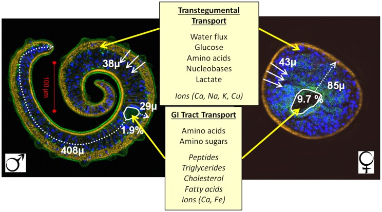
Schistosomes are parasitic flatworms that infect >200 million people worldwide, causing the chronic, debilitating disease schistosomiasis. Unusual among parasitic helminths, the long-lived adult worms, continuously bathed in blood, take up nutrients directly across the body surface and also by ingestion of blood into the gut. Recent proteomic analyses of the body surface revealed the presence of hydrolytic enzymes, solute, and ion transporters, thus emphasising its metabolic credentials. Furthermore, definition of the molecular mechanisms for the uptake of selected metabolites (glucose, certain amino acids, and water) establishes it as a vital site of nutrient acquisition. Nevertheless, the amount of blood ingested into the gut per day is considerable: for males ∼100 nl; for the more actively feeding females ∼900 nl, >4 times body volume. Ingested erythrocytes are lysed as they pass through the specialized esophagus, while leucocytes become tethered and disabled there. Proteomics and transcriptomics have revealed, in addition to gut proteases, an amino acid transporter in gut tissue and other hydrolases, ion, and lipid transporters in the lumen, implicating the gut as the site for acquisition of essential lipids and inorganic ions. The surface is the principal entry route for glucose, whereas the gut dominates amino acid acquisition, especially in females. Heme, a potentially toxic hemoglobin degradation product, accumulates in the gut and, since schistosomes lack an anus, must be expelled by the poorly understood process of regurgitation. Here we place the new observations on the proteome of body surface and gut, and the entry of different nutrient classes into schistosomes, into the context of older studies on worm composition and metabolism. We suggest that the balance between surface and gut in nutrition is determined by the constraints of solute diffusion imposed by differences in male and female worm morphology. Our conclusions have major implications for worm survival under immunological or pharmacological pressure.


Conflict of interest statement

The authors declare that no competing interests exist.

Figures

Figure 1
Figure 1. The tegument of adult S. mansoni.
Electron micrograph of a transverse section through the male tegument (T) and underlying musculature (M). The outer half is pitted (P, large arrowhead) extending the surface area, but notably lacks the microvillus-like microtriches of tapeworms, which acquire nutrients only across the tegument. The basal plasma membrane shows numerous infoldings (I, small arrowheads) typical of transporting epithelia. The cell bodies that contain the biosynthetic machinery lie below the muscle layer and are joined to the syncytium by narrow microtubule-lined cytoplasmic connections (CC, arrows). The inset shows the tegument surface at higher magnification revealing the two closely apposed lipid bilayers comprising an inner plasma membrane and an outer membranocalyx. The latter originates as the secreted contents of multilaminate vesicles (V, white arrows) that are produced by the Golgi apparatus in the cell bodies. Scale bars = 1 µm.
Figure 2
Figure 2. Layout of the alimentary tract.
A. Male S. haematobium showing the distribution of black hemozoin pigment that delineates the lumen of the gut caecum (c). The mouth at the base of the oral sucker (os) opens onto a short esophagus (es, arrows) that empties into an initial transverse region of gut (tg, arrowhead). This bifurcates to pass round the testes (te) before reuniting approximately halfway down the body to continue to the extreme posterior where it ends blindly. B. Female S. japonicum from a rabbit with the gut lumen almost completely filled with dark hemozoin pigment. The layout is the same as for the male but with the bifurcated caeca (c) passing first around the egg-filled uterus (ut, arrows) and ovary (o), before uniting to form a single tube completely surrounded by vitelline follicles (inset, higher magnification, esophageal region). C. Confocal image of the anterior of a male S. japonicum from a rabbit host highlighting the esophageal gland (green), revealed by detection of esophageal-specific protein SjMEG-4.1, and the nuclei (false-colored orange) stained by DAPI. The short esophagus is lined with atypical tegument syncytium, the surface of the anterior compartment (a) being corrugated while that in the posterior (p), coincident with the gland, is extended ∼50-fold by thin plate-like extensions. Aggregates of host leucocytes (leu, arrow) are evident in the esophageal lumen. Scale bars: A, 0.75 mm mm; B, 0.5 mm (inset, 0.2 mm); C, 0.1 mm.
Figure 3
Figure 3. The gut and protonephridia.
A. Transmission electron micrograph of the gut epithelium of an S. mansoni male. The cytoplasm of the syncytial gastrodermal epithelium (ga) is rich in rough endoplasmic reticulum (rer) and Golgi apparatus, typical of a cell synthesizing proteins and glycans for export; unlike the tegument it lacks obvious secretory inclusions. The luminal surface is extended by numerous thin lamellae (l) 3–5 microns long, in place of the conventional microvilli of an absorptive gut. Dense aggregates (“blobs”) of erythrocyte stroma (s) lie adjacent to the lamellae, together with paler lipid droplets (d). Inset, Stromal blob containing a hemozoin (Hz) pigment granule (g), with several more free granules adjacent. P, G and L denote Parenchyma, Gastrodermis and Lumen, respectively. B. Some (but not all) lipid droplets (d) have a dark ring of Hz around the periphery. C. The distribution of lipid droplets in the gut lumen, the epithelial syncytium and the surrounding parenchyma, is suggestive of transcytosis (i.e., the process of metabolite import into vesicles on one side of a cell followed by their release on the other side). D. Video frame from a feeding experiment during which an adult male S. mansoni regurgitated gut contents. Dark Hz demarcates the bifurcated gut (arrowheads) while a thin line of Hz (arrows) can be seen passing up the lumen of the posterior esophagus. (Numerous out-of-focus erythrocytes surround the worm. Dotted outline denotes ventral sucker.) E. Dorsal aspect of a female S. japonicum worm stained with FITC-labelled pea-nut agglutinin showing the bilateral distribution of flame cells (green dots) and protonephridial tubules running towards the main lateral collecting ducts on either side of the body. F. Higher magnification showing that each tubule terminates in a flame cell. The green dots are intense aggregates of O-glycan at the point where the flame cell connects to its tubule. Scale bars: A, 2 µm, inset 1 µm; B, C 0.4 µm; D, E 100 µm, F, 20 µm.
Figure 4
Figure 4. Pathways for diffusion of nutrients in male and female schistosomes.
Confocal images of stained adult male (left) and female (right) schistosome cross sections at the same magnification (yellow, phalloidin for actin; blue, DAPI for nuclei; green, tegument and gut). The parameter values are the mean of 12 females and 11 males in near perfect cross section. Areas and distances were measured using the polygon line and path tools in the Analyzing Digital Images package from the Lawrence Hall of Science, Berkeley, California (http://www.globalsystemsscience.org/software/download). Solid white arrows show diffusion distances from the tegument surface to the midpoint, dotted white arrows those from the gut lumen to the furthest extremity. The gut lumen of the female occupies 9.7% of the cross section, that of the male only 1.9%. Nutrients diffusing from the male gut would have to travel >400 µm to reach the furthest tissues whereas the maximum distance from the female gut is only 85 µm. The distance a nutrient has to diffuse from the tegument surface to the tissue midpoint in both male and female is very similar at ∼40 µm. Central boxes list metabolite classes that are transported across the tegument (upper box, yellow arrows) or via the gastrointestinal tract (lower box, yellow arrows). Metabolites that have been shown experimentally to be transported are listed in the upper group in each box. Those that have been inferred to be transported from proteomic, transcriptomic, or other work are indicated by italics.

Similar articles

Cited by

References

    1. Cheng TC, Bier JW (1972) Studies on molluscan schistosomiasis: an analysis of the development of the cercaria of Schistosoma mansoni. Parasitology 64: 129–141. - PubMed
    1. Hockley DJ (1972) Schistosoma mansoni: the development of the cercarial tegument. Parasitology 64: 245–252. - PubMed
    1. McLaren DJ, Hockley DJ (1977) Blood flukes have a double outer membrane. Nature 269: 147–149. - PubMed
    1. Fripp PJ (1967) The sites of (1-14C) glucose assimilation in Schistosoma haematobium. Comp Biochem Physiol 23: 893–898. - PubMed
    1. Skelly PJ, Tielens AGM, Shoemaker CB (1998) Glucose transport and metabolism in mammalian stage schistosomes. Parasitol Today 14: 402–406. - PubMed

Publication types

LinkOut - more resources