. 2014 Aug 14;10(8):e1004505.
doi: 10.1371/journal.pgen.1004505. eCollection 2014 Aug.

Conditional inactivation of Upstream Binding Factor reveals its epigenetic functions and the existence of a somatic nucleolar precursor body

Affiliations

Conditional inactivation of Upstream Binding Factor reveals its epigenetic functions and the existence of a somatic nucleolar precursor body

Nourdine Hamdane et al. PLoS Genet. .

Abstract

Upstream Binding Factor (UBF) is a unique multi-HMGB-box protein first identified as a co-factor in RNA polymerase I (RPI/PolI) transcription. However, its poor DNA sequence selectivity and its ability to generate nucleosome-like nucleoprotein complexes suggest a more generalized role in chromatin structure. We previously showed that extensive depletion of UBF reduced the number of actively transcribed ribosomal RNA (rRNA) genes, but had little effect on rRNA synthesis rates or cell proliferation, leaving open the question of its requirement for RPI transcription. Using gene deletion in mouse, we now show that UBF is essential for embryo development beyond morula. Conditional deletion in cell cultures reveals that UBF is also essential for transcription of the rRNA genes and that it defines the active chromatin conformation of both gene and enhancer sequences. Loss of UBF prevents formation of the SL1/TIF1B pre-initiation complex and recruitment of the RPI-Rrn3/TIF1A complex. It is also accompanied by recruitment of H3K9me3, canonical histone H1 and HP1α, but not by de novo DNA methylation. Further, genes retain penta-acetyl H4 and H2A.Z, suggesting that even in the absence of UBF the rRNA genes can maintain a potentially active state. In contrast to canonical histone H1, binding of H1.4 is dependent on UBF, strongly suggesting that it plays a positive role in gene activity. Unexpectedly, arrest of rRNA synthesis does not suppress transcription of the 5S, tRNA or snRNA genes, nor expression of the several hundred mRNA genes implicated in ribosome biogenesis. Thus, rRNA gene activity does not coordinate global gene expression for ribosome biogenesis. Loss of UBF also unexpectedly induced the formation in cells of a large sub-nuclear structure resembling the nucleolar precursor body (NPB) of oocytes and early embryos. These somatic NPBs contain rRNA synthesis and processing factors but do not associate with the rRNA gene loci (NORs).


Conflict of interest statement

The authors have declared that no competing interests exist.

Figures
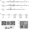
Figure 1
Figure 1. The Ubf gene is essential for mouse development beyond morula.
A) Structure of the wild type Ubf (WT), the conditional Ubf−fl (Floxed), and the deleted Ubf −Δ (Δ) alleles. B) Survival statistics for Ubfwt/wt, Ubf Δ/wt and Ubf Δ/Δ mouse embryos and offspring. Note that no embryos were detected at or after 8.5 dpc. C) Examples of mouse embryos and genotyping at the equivalent of 3.5 dpc. Ubf null embryos arrest at the morula stage. D) In vitro development of 3.5 dpc embryos to late blastocysts (equivalent to 4.5 dpc). Ubf null embryos do not develop further and necrose.
Figure 2
Figure 2. UBF is essential for the synthesis of the major rRNAs in cell culture.
A) to E) Ubffl/fl/Er-cre+/+ and Ubfwt/wt Er-cre+/+ cells were treated with 4-HT to induce recombination in the Ubf gene and at the indicated time points, A) genotyped for Ubf recombination, B) analyzed by Western blot for UBF levels, and C) metabolically labelled with [3H]-uridine to follow rRNA synthesis of the rRNAs and their precursors. D) Northern blot analysis of the 47S and 34S rRNA pools. The upper diagram shows the organisation of the larger rRNA precursors and the probe used. In C) “Bulk” refers to the EtBr stained total RNA fractionation. E) Quantitative analyses of rRNA synthesis rates and pool sizes.
Figure 3
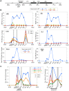
Figure 3. UBF elimination causes release of the RPI and all RPI initiation factors, but not of the termination factor TTF1.
A) Map of the rDNA repeat showing above; the coding and Enhancer regions, and below; the amplicons sampled by ChIP analysis. B) Association of UBF, RPI, TTF1, SL1 (TIF1B) and Rrn3 (TIF1A) and of initiation competent RPI (Rrn3/RPI) determined by ChIP/Q-PCR assays of chromatin from Ubffl/fl/Er-cre+/+ cells prepared at the indicated times pHT. C) Relative distribution of RPI, SL1, Rrn3 and UBF at 0 h pHT, i.e. before Ubf recombination. Left panel shows data on an enlarged horizontal scale. In B and C the horizontal axes indicate the position of amplicons, and the grey bars the positions of known binding sites for TTF1. Ubf gene recombination and UBF protein levels were assayed in parallel with ChIP analyses and followed those shown in Figure 2A and B. The data shown in B and C are given after subtraction of the parallel preimmune Control ChIP data. They are derived from single ChIP preparations analyzed in triplicate, but are representative of the data from biological replicates, see Materials and Methods.
Figure 4
Figure 4. The rDNA chromatin is extensively remodelled during UBF elimination.
A) Psoralen crosslinking analysis of the rRNA genes in Ubffl/fl/Er-cre+/+ and Ubfwt/wt/Er-cre+/+ cells at the indicated times pHT. The probe position is indicated in the upper diagram, and the lower panel shows a typical electrophoretic separation of actively transcribed “a” and inactive “i” genes. The right-hand panel displays a quantitation of the fraction of transcriptionally active and inactive genes at each time point. Elimination of UBF at 48 h pHT correlates with full inactivation of the rRNA genes. B) and C) Changes in histone modifications penta-acetyl H4 (H4ac) and H3K9me3, and relative Histone H1, H2AZ, H1.4, H1.4-S187p and chromosomal protein HP1α levels associated with the rDNA during UBF elimination (hours pHT). The grey band indicates the extent of the rRNA gene Enhancer sequences. Mapping of amplicons is as in Figure 3A. Again here Ubf gene recombination and UBF protein levels were assayed in parallel and closely followed those shown in Figure 2A and B. The data shown in B and C are given after subtraction of the parallel preimmune Control ChIP data. They are derived from single ChIP preparations analyzed in triplicate, but are representative of the data from biological replicates, see Materials and Methods.
Figure 5
Figure 5. UBF elimination has only minor effects on global gene expression.
A) Metabolic labeling of Ubffl/fl/Er-cre+/+ and Ubfwt/wt/Er-cre+/+ cells with [3H]-uridine to follow synthesis of U2 and U3 snRNAs, 5.8S rRNA, 5S rRNA, tRNAs and 18S at the given time points pHT. “Bulk 18S” refers to the EtBr input control. The ratio of incorporation into 5.8S relative to 5S (5.8S/5S) is given below each track of the upper panel. B) Boxplot of unbiased expression microarray data for various gene classes. The plots indicate the relative gene expression levels for Ubffl/fl/Er-cre+/+ cells at 72 h pHT relative to levels for Ubfwt/wt/Er-cre+/+ at the same time point. The original data can be found in the GEO databank under the accession number GSE55450.
Figure 6
Figure 6. UBF elimination reveals that nucleolar protein bodies exist independently of rRNA gene activity.
A) to C) 3D IF analysis of respectively UBF, RPI (large subunit, A194) and Rrn3/TIF1A relative to Fibrillarin and DNA stained with DAPI. The subnuclear volumes of the indicated proteins are indicated as surfaces of constant fluorescence intensity (isosurface) with directional pseudo-illumination to indicate their 3D form. D) Left; quantitation of the mean number of (Fibrillarin positive) nucleoli or nucleolar bodies per nucleus, and right; mean (Fibrillarin) volume of individual nucleoli. Data were the mean of three independent series of IF analyses, and ∼20 nuclei were analyzed per time point.
Figure 7
Figure 7. Somatic Nucleolar Precursor Bodies are distinguished from nucleoli by not being visible in bright field.
IF and bright field images of Ubffl/fl/Er-cre+/+ cells at 0 and 72 h pHT counterstained with DAPI and showing the location of RPI (red) and fibrillarin (green).
Figure 8
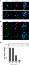
Figure 8. Nucleolar bodies are spatially distinct from the rDNA and NORs.
A) and B) 3D Immuno-FISH analysis of the spatial distribution of rDNA relative to Fibrillarin and TTF1 at the indicated times pHT treatment. The subnuclear volumes of the indicated proteins and DNA are indicated as surfaces of constant fluorescence intensity (isosurface) with directional pseudo-illumination to indicate their 3D form. C) Quantitative analysis of the 3D spatial correlation of rDNA FISH fluorescence with Fibrillarin and with TTF1 fluorescence. Data were the mean of two independent Immuno-FISH analyses in which >20 nuclei were analyzed.
Figure 9
Figure 9. A model for the chromatin structure of the three distinct states of rRNA gene activity.
Active, unmethylated (meCpG minus) genes maintain UBF and phospho-H1.4 (H1.4p) over their Enhancer region and their 47S transcribed gene region (indicated by lateral fibrils and rRNA gene blocks), while RPI is restricted to the 47S region and to a site close to the spacer terminator (Tsp), the latter probably in an arrested elongation state. These same gene regions display variable levels of H4ac, and are denuded of nucleosomes. H2Az is most likely present adjacent to the spacer promoter (SpPr), while the preinitiation complex SL1/TIF1B is present at both spacer and 47S Promoters (Pr), (not shown). The Inactive, unmethylated (meCpG minus) fraction of genes is devoid of UBF, displays enhanced occupation by H3K9me3 and HP1α, especially towards the 3′ 47S region, and nucleosomes replace UBF over most of its interaction domain. But these inactive genes retain H2Az and H4ac over the SpPr and Enhancer regions, suggesting they are in dynamic exchange with the actively transcribed gene population. Our unpublished alignments of H3K4me3 and CTCF Encode data sets for ES and MEFs show that these factors are also present flanking the SpPr probably on both Active and Inactive unmethylated genes. The methylated (meCpG) gene fraction is unaffected by UBF loss, and most probably exists in a classical heterochromatic state corresponding to the inherited constitutively silenced NORs. Due to limited data, the status of the “Enhancer” region is difficult to ascertain and this is indicated by a different shading.

References

    1. Derenzini M, Trerè D, Pession A, Montanaro L, Sirri V, et al. (1998) Nucleolar function and size in cancer cells. AmJPathol 152: 1291–1297. - PMC - PubMed
    1. Vlatkovic N, Boyd MT, Rubbi CP (2013) Nucleolar control of p53: a cellular Achilles' heel and a target for cancer therapy. Cell Mol Life Sci 71: 771–791. - PMC - PubMed
    1. Moss T, Langlois F, Gagnon-Kugler T, Stefanovsky V (2007) A housekeeper with power of attorney: the rRNA genes in ribosome biogenesis. Cell Mol Life Sci 64: 29–49. - PMC - PubMed
    1. Bywater MJ, Pearson RB, McArthur GA, Hannan RD (2013) Dysregulation of the basal RNA polymerase transcription apparatus in cancer. Nat Rev Cancer 13: 299–314. - PubMed
    1. Lessard F, Morin F, Ivanchuk S, Langlois F, Stefanovsky V, et al. (2010) The ARF tumor suppressor controls ribosome biogenesis by regulating the RNA polymerase I transcription factor TTF-I. Mol Cell 38: 539–550. - PubMed

Publication types

MeSH terms

Associated data