. 2014 Oct;28(10):891-902.
doi: 10.1177/0269881114543719. Epub 2014 Aug 13.

Vortioxetine disinhibits pyramidal cell function and enhances synaptic plasticity in the rat hippocampus

Affiliations

Vortioxetine disinhibits pyramidal cell function and enhances synaptic plasticity in the rat hippocampus

Elena Dale et al. J Psychopharmacol. 2014 Oct.

Erratum in

  • J Psychopharmacol. 2014 Dec;28(12):1192

Abstract

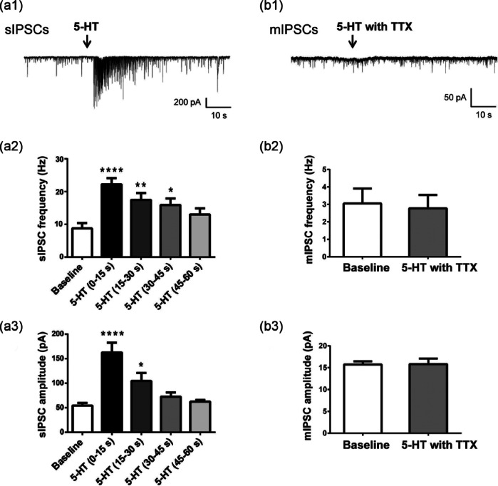
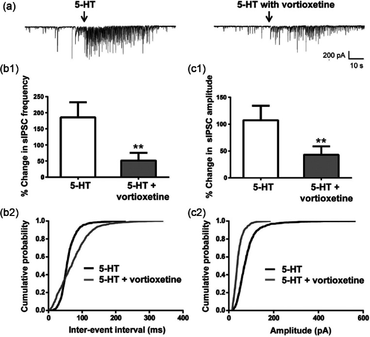
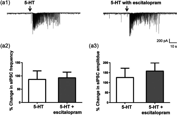
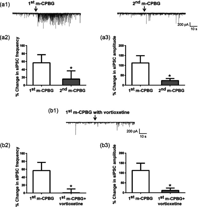
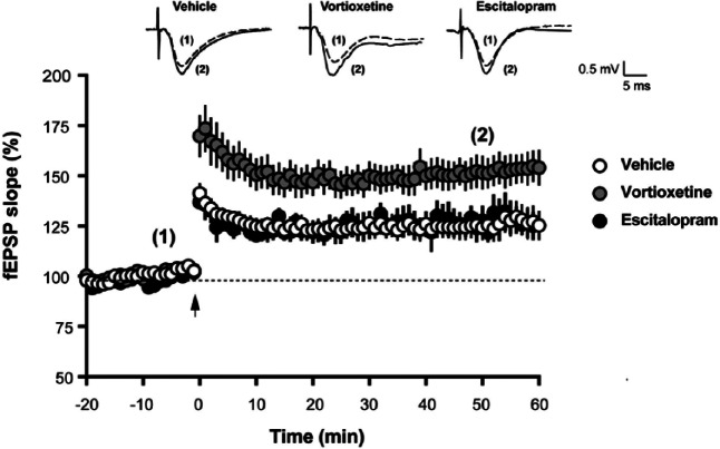
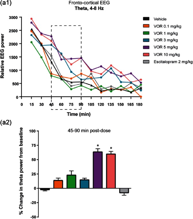
Vortioxetine, a novel antidepressant with multimodal action, is a serotonin (5-HT)3, 5-HT7 and 5-HT1D receptor antagonist, a 5-HT1B receptor partial agonist, a 5-HT1A receptor agonist and a 5-HT transporter (SERT) inhibitor. Vortioxetine has been shown to improve cognitive performance in several preclinical rat models and in patients with major depressive disorder. Here we investigated the mechanistic basis for these effects by studying the effect of vortioxetine on synaptic transmission, long-term potentiation (LTP), a cellular correlate of learning and memory, and theta oscillations in the rat hippocampus and frontal cortex. Vortioxetine was found to prevent the 5-HT-induced increase in inhibitory post-synaptic potentials recorded from CA1 pyramidal cells, most likely by 5-HT3 receptor antagonism. Vortioxetine also enhanced LTP in the CA1 region of the hippocampus. Finally, vortioxetine increased fronto-cortical theta power during active wake in whole animal electroencephalographic recordings. In comparison, the selective SERT inhibitor escitalopram showed no effect on any of these measures. Taken together, our results indicate that vortioxetine can increase pyramidal cell output, which leads to enhanced synaptic plasticity in the hippocampus. Given the central role of the hippocampus in cognition, these findings may provide a cellular correlate to the observed preclinical and clinical cognition-enhancing effects of vortioxetine.

Keywords: 5-hydroxytryptamine 3 receptor; CA1; Serotonin; cognition; electroencephalography; long-term potentiation.


Conflict of interest statement

E Dale, H Zhang, SC Leiser, N Plath and C Sanchez are full-time employees of Lundbeck. The work of Y Xiao, D Lu and C Yang was sponsored by H Lundbeck A/S.

Figures

Figure 1.
Figure 1.
Serotonin (5-HT) increased frequency and amplitude of spontaneous inhibitory post-synaptic currents (sIPSCs), but not miniature inhibitory post-synaptic currents (mIPSCs), recorded from hippocampal CA1 pyramidal cells. (a1), (b1) Representative traces of sIPSCs (a1) and mIPSCs (b1) recorded from a CA1 pyramidal cell before and after local application of 5-HT (100 µM for 500 ms). mIPSCs were recorded in the presence of 1 µM tetrodotoxin (TTX). (a2), (a3) Serotonin (5-HT) transiently increased the frequency ((a2), ****p<0.001, **p=0.0078, *p=0.0361, one-way analysis of variance (ANOVA) with Dunnett’s multiple comparisons test vs baseline) and amplitude ((a3), ****p<0.001, *p=0.0235, one-way ANOVA with Dunnett’s multiple comparisons test vs baseline) of sIPSCs. The largest increase was observed in the first 15 s after 5-HT application and the response to 5-HT was largely desensitized after 60 s. Bars represent the mean±standard error of the mean (SEM) of recordings from 10 cells. (b2), (b3) 5-HT had no effect on mIPSC frequency (b2) or amplitude (b3) (p>0.05, paired Student’s t-test). Bars represent the mean±SEM of 60 s recordings from four cells.
Figure 2.
Figure 2.
Vortioxetine blocked the serotonin (5-HT)-induced increase in spontaneous inhibitory post-synaptic currents (sIPSCs). (a) Representative sIPSCs recorded from a CA1 pyramidal neuron in response to 5-HT before (left) and after 15 min application of 20 µM vortioxetine (right). (b1), (c1) Vortioxetine blocked the 5-HT induced increase in sIPSC frequency ((b1), **p=0.0073, paired Student’s t-test) and amplitude ((c1), **p=0.0066, paired Student’s t-test). Frequency and amplitude were normalized to the mean value during the 30 s of recordings prior to 5-HT application. Bars represent the mean±standard error of the mean (SEM) of sIPSCs from 15 cells. (b2), (c2) Averaged cumulative probability plots of sIPSC inter-event interval (b2) and amplitude (c2) for 5-HT alone and 5-HT+vortioxetine. Data for these graphs were derived from 60 s of continuous recordings. Each data point represents the mean from 15 cells. Vortioxetine significantly increased the inter-event interval time in 14/15 cells and shifted amplitudes of sIPSCs to lower values in 13/15 cells after 5-HT application (p<0.05, Kolmogorov-Smirnov (K-S) test).
Figure 3.
Figure 3.
Escitalopram had no effect on serotonin (5-HT) increase of spontaneous inhibitory post-synaptic currents (sIPSCs). (a1) Representative sIPSCs recorded from a CA1 pyramidal neuron in response to 5-HT before (left) and after 15 min perfusion with 10 µM escitalopram (right). (a2), (a3) Escitalopram did not change the 5-HT response on sIPSC frequency (a2) or amplitude (a3) (p>0.05, paired Student’s t-test). Frequency and amplitude were normalized to the mean value during the 30 s of recordings prior to 5-HT application. Bar graphs represent the mean±standard error of the mean (SEM) from six cells.
Figure 4.
Figure 4.
The serotonin (5-HT)3 receptor agonist m-chlorophenylbiguanide hydrochloride (m-CPBG) transiently increased spontaneous inhibitory post-synaptic currents (sIPSCs) and vortioxetine blocked the m-CPBG effect. (a1) Representative sIPSCs recorded from a CA1 pyramidal neuron in response to two applications of the 5-HT3 receptor agonist m-CPBG. In most of the responding cells, the effect of m-CPBG was not repeatable after various application intervals (compare right and left traces). (a2), (a3) The second application of m-CPBG did not induce the same increase in sIPSC frequency ((a2), *p=0.031, paired Student’s t-test) or amplitude ((a3), *p=0.024, paired Student’s t-test) as the first application. Bar graphs represent the mean±standard error of the mean (SEM) from 11 cells. Because of strong desensitization of m-CPBG responses, the effect of vortioxetine was tested in a separate set of cells. (b1) Representative sIPSCs recorded from a CA1 pyramidal neuron in response to m-CPBG in the presence of 20 µM vortioxetine. (b2), (b3) Pre-treatment with vortioxetine blocked the m-CPBG increase in sIPSC frequency ((b2), *p=0.034, unpaired Student’s t-test) and amplitude ((b3), *p=0.028, unpaired Student’s t-test). Frequency and amplitude were normalized to the mean value during the 30 s of recordings prior to 5-HT application. Bar graphs represent the mean±SEM from 11 cells for the m-CPBG only condition and from nine cells for the m-CPBG + vortioxetine condition.
Figure 5.
Figure 5.
Vortioxetine enhanced theta burst long-term potentiation (LTP) in hippocampal slices. Representative traces of field excitatory post-synaptic potential (fEPSP) recordings from vehicle-treated, vortioxetine-treated and escitalopram-treated slices are shown on top of the graph. Each trace is the mean of five sweeps taken either immediately before (dashed line marked with (1) or 50 min after (solid line marked with (2)) theta-burst stimulation (TBS). TBS, marked with an arrow in the time course graph, induced a long-lasting increase in the slopes of fEPSPs. For each time point, fEPSP slopes were calculated from either vehicle-treated (n=17 slices from 12 animals), vortioxetine-treated (n=14 from nine animals) or escitalopram-treated slices (n=5 slices from four animals) and expressed as % of baseline. Data are shown as the mean±standard error of the mean (SEM). Perfusion of hippocampal slices with vortioxetine for 30 min prior to TBS increased LTP without affecting baseline transmission (p=0.0017 vortioxetine vs vehicle, two-way analysis of variance (ANOVA) F(2,38)=7.548)). Escitalopram had no effect on LTP (p>0.05).
Figure 6.
Figure 6.
Vortioxetine increased frontal cortical theta power in in vivo electroencephalography (EEG) recordings. (a1) Total relative theta power are shown for each treatment for every 15 min from 0–180 min postdose. A treatment effect was observed for vortioxetine (VOR), but not escitalopram (p=0.0226, one-way analysis of variance (ANOVA) F(65,497)=1.4180). (a2) Pharmacologically-induced changes in EEG recordings were quantified by averaging the 10 s bins constituting 15–75 min before (baseline) and 45–90 min post-drug treatment. Theta power was expressed as the percentage change from baseline. Bar graphs represent the mean±standard error of the mean (SEM) (n=9 rats per condition). A negative value indicates a decrease in EEG power from baseline. Vortioxetine at 5.0 and 10 mg/kg significantly increased theta power (*p<0.05, post-dose versus vehicle, one-way analysis of variance (ANOVA) with least significant difference (LSD) post-hoc comparison). Escitalopram at 2.0 mg/kg had no effect on theta power.

References

    1. Adell A. (2010) Lu-AA21004, a multimodal serotonergic agent, for the potential treatment of depression and anxiety. IDrugs 13: 900–910. - PubMed
    1. Alvarez E, Perez V, Dragheim M, et al. (2012) A double-blind, randomized, placebo-controlled, active reference study of Lu AA21004 in patients with major depressive disorder. Int J Neuropsychopharmacol 15: 589–600. - PMC - PubMed
    1. Andrade R, Nicoll RA. (1987) Pharmacologically distinct actions of serotonin on single pyramidal neurones of the rat hippocampus recorded in vitro. J Physiol 394: 99–124. - PMC - PubMed
    1. Arai A, Lynch G. (1992) Factors regulating the magnitude of long-term potentiation induced by theta pattern stimulation. Brain Res 598: 173–184. - PubMed
    1. Arnsten AF, Lin CH, Van Dyck CH, et al. (1997) The effects of 5-HT3 receptor antagonists on cognitive performance in aged monkeys. Neurobiol Aging 18: 21–28. - PubMed

Publication types

MeSH terms