. 2014 Aug;26(8):3314-25.
doi: 10.1105/tpc.114.129171. Epub 2014 Aug 8.

Identification of a sphingolipid α-glucuronosyltransferase that is essential for pollen function in Arabidopsis

Affiliations

Identification of a sphingolipid α-glucuronosyltransferase that is essential for pollen function in Arabidopsis

Emilie A Rennie et al. Plant Cell. 2014 Aug.

Abstract

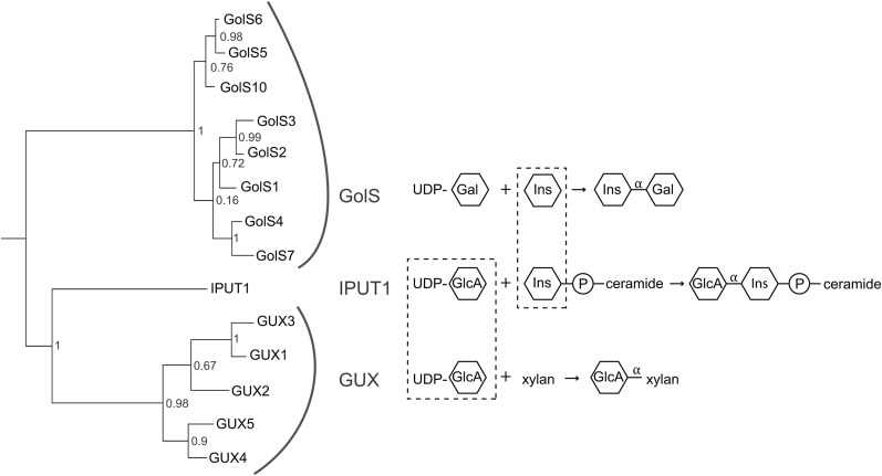
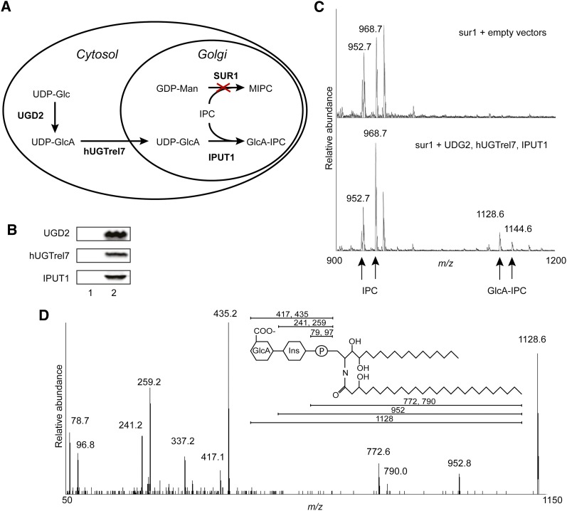
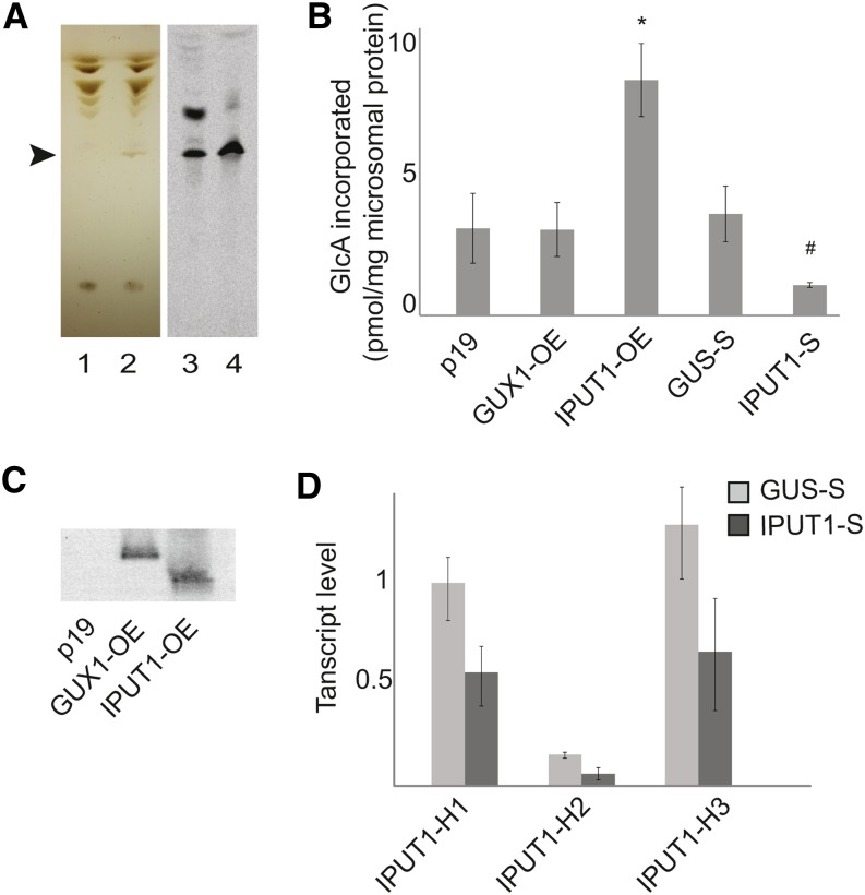
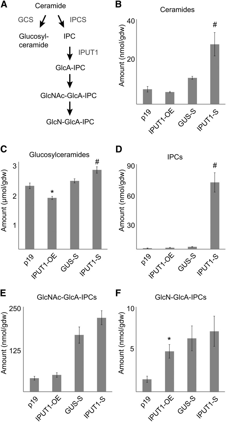
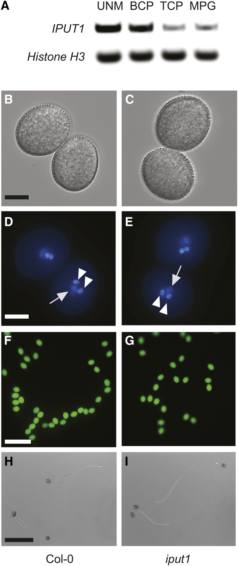
Glycosyl inositol phosphorylceramide (GIPC) sphingolipids are a major class of lipids in fungi, protozoans, and plants. GIPCs are abundant in the plasma membrane in plants, comprising around a quarter of the total lipids in these membranes. Plant GIPCs contain unique glycan decorations that include a conserved glucuronic acid (GlcA) residue and various additional sugars; however, no proteins responsible for glycosylating GIPCs have been identified to date. Here, we show that the Arabidopsis thaliana protein INOSITOL PHOSPHORYLCERAMIDE GLUCURONOSYLTRANSFERASE1 (IPUT1) transfers GlcA from UDP-GlcA to GIPCs. To demonstrate IPUT1 activity, we introduced the IPUT1 gene together with genes for a UDP-glucose dehydrogenase from Arabidopsis and a human UDP-GlcA transporter into a yeast mutant deficient in the endogenous inositol phosphorylceramide (IPC) mannosyltransferase. In this engineered yeast strain, IPUT1 transferred GlcA to IPC. Overexpression or silencing of IPUT1 in Nicotiana benthamiana resulted in an increase or a decrease, respectively, in IPC glucuronosyltransferase activity in vitro. Plants in which IPUT1 was silenced accumulated IPC, the immediate precursor, as well as ceramides and glucosylceramides. Plants overexpressing IPUT1 showed an increased content of GIPCs. Mutations in IPUT1 are not transmitted through pollen, indicating that these sphingolipids are essential in plants.


Figures

Figure 1.
Figure 1.
Phylogenetic Relationship of the GolS, GUX, and IPUT1 Proteins. IPUT1 is related to both the GolS and GUX proteins, which transfer sugars with a retaining mechanism that creates α glycosidic linkages. IPUT1 uses the same substrate, UDP-GlcA, as the GUX proteins, while using the same acceptor, inositol (Ins) or inositol phosphorylceramide, as the GolS proteins (dashed lines). Likelihood values are given at nodes.
Figure 2.
Figure 2.
Production of GlcA-IPC in Yeast. (A) Diagram of strategy for expressing proteins to produce GlcA-IPC. UDP-Glucose Dehydrogenase 2 (UGD2), human UDP-Galactose Transporter Relative 7 (hUGTrel7), and IPUT1 were transformed into sur1Δ yeast. Cross indicates deletion of sur1. (B) Immunoblot showing expression of heterologous proteins in yeast with empty vectors (1) or vectors driving expression of UGD2, hUGTrel7, and IPUT1 (2). (C) Mass spectrometry analysis of lipids from yeast transformed with control (empty) or UGD2, hUGTrel7, and IPUT1 vectors. Major IPC species at m/z 952.7 and 968.7 containing trihydroxy C18 saturated long-chain sphingobases acylated with mono- or dihydroxy C26 saturated fatty acids, respectively, and corresponding GlcA-IPC species at m/z 1128.6 and 1144.6 are indicated. (D) Tandem mass spectrometry confirming identification of the GlcA-IPC peak at m/z 1128.6. Expected m/z for each fragment ion is indicated in the diagram. [See online article for color version of this figure.]
Figure 3.
Figure 3.
Assay for IPC Glucuronosyltransferase Activity in N. benthamiana. (A) TLC of lipids extracted from sur1Δ yeast with empty vectors (1) or vectors with UGD2, hUGTrel7, and IPUT1 (2). GlcA-IPC is indicated by the arrowhead. Radiolabeled lipids from N. benthamiana microsomes incubated with UDP-[14C]GlcA followed by mock (3) or mild alkaline hydrolysis treatment (4) were developed on the same TLC plate. (B) Incorporation of radiolabel into GlcA-IPC bands produced from microsomes prepared from N. benthamiana plants expressing p19 (control), overexpressing GUX1 (GUX-OE) or IPUT1 (IPUT1-OE), or plants silencing the control gene GUS (GUS-S) or IPUT1 (IPUT1-S). *, Significantly different from p19 (t test, P < 0.05); #, significantly different from GUS-S (t test, P < 0.05). (C) Immunoblot of microsomal proteins from N. benthamiana plants expressing p19 or overexpressing GUX1 (GUX-OE) or IPUT1 (IPUT1-OE). (D) Expression levels of N. benthamiana IPUT1 homologs H1, H2, and H3 in IPUT1-silenced plants. All transcript levels were normalized to ACT2, UBI, and EF1α. Values shown are normalized to the transcript level of IPUT1-H1 in GUS-silenced plants. All values are the mean ± se of three biological replicates. [See online article for color version of this figure.]
Figure 4.
Figure 4.
Sphingolipid Profiles in N. benthamiana Plants with Altered Expression of IPUT1. (A) Outline of complex sphingolipid synthesis. Known enzymes in the pathway are shown in gray. GCS, glucosylceramide synthase. (B) to (F) Profiles of sphingolipids extracted from leaves of plants expressing p19 (control), overexpressing IPUT (IPUT1-OE), silencing the control gene (GUS-S), or silencing IPUT1 (IPUT1-S). *, Significantly different from p19 (t test, P < 0.05); #, significantly different from GUS-S (t test, P < 0.05). gdw, gram dry weight. Values are the mean ± se of five biological replicates.
Figure 5.
Figure 5.
iput1 Pollen Develops Normally. (A) RT-PCR of IPUT1 and Histone H3 (control) in the uninucleate microspore (UNM), bicellular pollen (BCP), tricellular pollen (TCP), and mature pollen grains (MPG). RT-PCRs used pooled spores from two biological replicates. (B), (D), (F), and (H) Pollen from wild-type plants. (C), (E), (G), and (I) Pollen from iput1-1/+ plants. Phenotype of wild type and iput1 pollen is indistinguishable. Light microscopy of mature pollen grains ([A] and [B]); fluorescence microscopy after DAPI staining ([D] and [E]) and fluorescein diacetate staining ([F] and [G]). Arrows indicate the diffusely stained vegetative nucleus; arrowheads indicate the densely stained sperm cell nuclei. Light microscopy of germinated pollen tubes, 6 h after germination ([H] and [I]). Bars = 10 μm in (B) to (F) and 100 μm in (G) to (I).

References

    1. Beeler T.J., Fu D., Rivera J., Monaghan E., Gable K., Dunn T.M. (1997). SUR1 (CSG1/BCL21), a gene necessary for growth of Saccharomyces cerevisiae in the presence of high Ca2+ concentrations at 37 ° C, is required for mannosylation of inositolphosphorylceramide. Mol. Gen. Genet. 255: 570–579. - PubMed
    1. Boavida L.C., McCormick S. (2007). Technical advance: temperature as a determinant factor for increased and reproducible in vitro pollen germination in Arabidopsis thaliana. Plant J. 52: 570–582. - PubMed
    1. Borner G.H.H., Sherrier D.J., Weimar T., Michaelson L.V., Hawkins N.D., Macaskill A., Napier J.A., Beale M.H., Lilley K.S., Dupree P. (2005). Analysis of detergent-resistant membranes in Arabidopsis. Evidence for plasma membrane lipid rafts. Plant Physiol. 137: 104–116. - PMC - PubMed
    1. Bromley P.E., Li Y.O., Murphy S.M., Sumner C.M., Lynch D.V. (2003). Complex sphingolipid synthesis in plants: characterization of inositolphosphorylceramide synthase activity in bean microsomes. Arch. Biochem. Biophys. 417: 219–226. - PubMed
    1. Brownfield L., Hafidh S., Borg M., Sidorova A., Mori T., Twell D. (2009). A plant germline-specific integrator of sperm specification and cell cycle progression. PLoS Genet. 5: e1000430. - PMC - PubMed

Publication types

MeSH terms