. 2014 Sep;9(9):2135-46.
doi: 10.1038/nprot.2014.143. Epub 2014 Aug 14.

Addition of exogenous α-synuclein preformed fibrils to primary neuronal cultures to seed recruitment of endogenous α-synuclein to Lewy body and Lewy neurite-like aggregates

Affiliations

Addition of exogenous α-synuclein preformed fibrils to primary neuronal cultures to seed recruitment of endogenous α-synuclein to Lewy body and Lewy neurite-like aggregates

Laura A Volpicelli-Daley et al. Nat Protoc. 2014 Sep.

Abstract

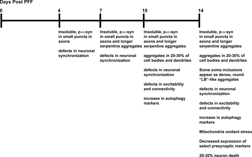
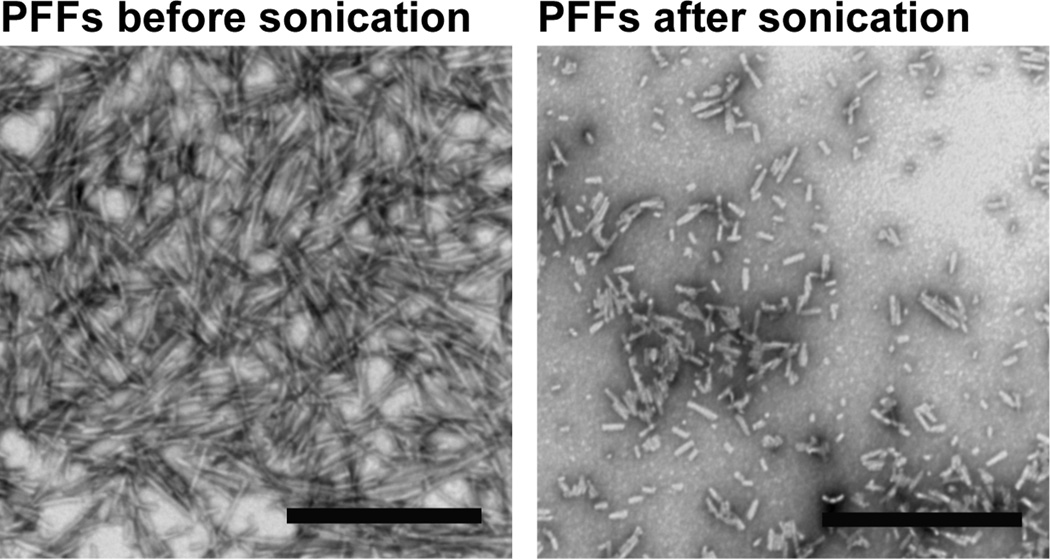
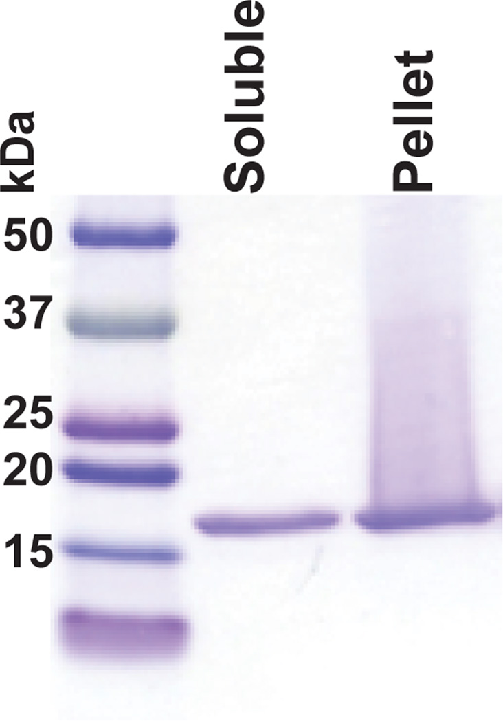
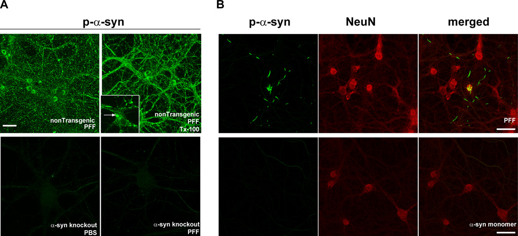
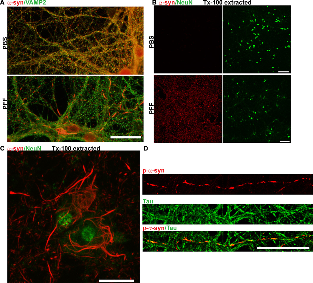
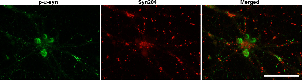
This protocol describes a primary neuronal model of formation of α-synuclein (α-syn) aggregates that recapitulate features of the Lewy bodies and Lewy neurites found in Parkinson's disease brains and other synucleinopathies. This model allows investigation of aggregate formation, their impact on neuron function, and development of therapeutics. Addition of preformed fibrils (PFFs) synthesized from recombinant α-syn to neurons seeds the recruitment of endogenous α-syn into aggregates characterized by detergent insolubility and hyperphosphorylation. Aggregate formation follows a lag phase of 2-3 d, followed by formation in axons by days 4-7, spread to somatodendritic compartments by days 7-10 and neuron death ~14 d after PFF addition. Here we provide methods and highlight the crucial steps for PFF formation, PFF addition to cultured hippocampal neurons and confirmation of aggregate formation. Neurons derived from various brain regions from nontransgenic and genetically engineered mice and rats can be used, allowing interrogation of the effect of specific genes on aggregate formation.


Figures

Figure 1
Figure 1
Timeline of known events in PFF model of α-syn aggregation.
Figure 2
Figure 2
Electron micrographs of PFFs before (A) and after (B) sonication. Scale bars= 500 nm.
Figure 3
Figure 3. Typical results seen at step 12 following the sedimentation assay of PFFs
PFFs were prepared as described in steps 1–12 and the supernatant and pellet resolved by SDS-PAGE and stained with Coomassie Blue. This batch of PFFs had approximately equivalent amounts of α-syn in the supernatant and pellet was efficient at seeding aggregates from endogenous α-syn in primary neurons.
Figure 4
Figure 4. Visualization of PFF-induced formation of α-syn aggregates using an antibody to p-α-syn
A. Fourteen days following the addition of PFFs to nontransgenic primary neurons, p-α-syn inclusions, visualized by immunofluorescence using mAB81A, were abundant throughout neurites and soma (top panels). These inclusions were also insoluble as determined by fixation in paraformaldehyde/4% sucrose/1% Tx (top, right). p-α-Syn inclusions were not apparent in PBS or PFF treated (14 days post-treatment) in primary neurons from α-syn knockout mice (bottom panels). Scale bar = 50 µm. B. PFFs (top panels) or monomeric, non-fibrillar α-syn (bottom panels) were added to primary neurons. Immunofluorescent imaging was performed using antibodies MJFR1, which recognizes p-α-syn, and NeuN, to visualize neuronal soma. Inclusions positive for p-α-syn (green) were abundant in PFF-treated neurons, but not neurons treated with soluble, monomeric α-syn. Scale bar = 20 µm.
Figure 5
Figure 5. Immunofluorescence of α -syn aggregates
A. Neurons were fixed in 4% paraformaldehyde/4% sucrose and immunofluorescence was performed using an antibody to total α-syn (Syn1) and an antibody which recognizes the presynaptic marker, VAMP2 In control PBS-treated neurons, α-syn colocalized extensively with VAMP2 at presynaptic terminals. Fourteen days following PFF treatment, α-syn no longer localized to the presynaptic terminal, but was found in longer serpentine aggregates in axons and skein-like filaments in the soma. Scale bar = 10 µm. B. Neurons were fixed in 4% paraformaldehyde/4% sucrose/1% Tx-100 and immunofluorescence was performed using an antibody to total α-syn and an antibody which recognizes the neuron specific marker, NeuN. In control neurons, α-syn was completely extractable but in PFF treated neurons (here, 10 days post), α-syn was insoluble and thus visible throughout the culture. Scale bar = 50 µm. C. 4% paraformaldehyde/4% sucrose/1% Tx-100 and co-stained using an antibody to total α-syn and NeuN. Scale bar = 10 µm. D. Seven days following PFF treatment, neurons were fixed with 4% paraformaldehyde/4% sucrose and co-stained with a p- α-syn (red) and tau (green) antibody to label axons.Scale bar = 10 µm.
Figure 6
Figure 6. Visualization of exogenously added PFFs and p-α-syn aggregates formed from endogenous α-syn
Neurons were fixed 14 days after treatment with PFFs and double immunofluorescence was performed using a rabbit antibody to p-α-syn generated in the Lee lab (green), and a mouse antibody, Syn204, that specifically recognizes the exogenously added human PFFs (red). There was minimal colocalization between the exogenous PFFs and the p-α-syn inclusions derived from endogenous α-syn. Scale bar = 50 µm.
Figure 7
Figure 7. Typical immunoblot results seen following sequential extraction of neurons in 1% Tx-100 followed by SDS
A. Immunoblotting was performed using Syn1, an antibody that recognizes total α-syn. In control neurons, α-syn was completely extractable in Tx-100. In PFF treated neurons, there was a decrease in Tx-100 soluble α-syn, and increase in SDS-soluble α-syn. B. Immunoblots of SDS extracts (Tx-100 insoluble) using mAB81a that recognizes p-α-syn. There was no p-α-syn in control neurons but a substantial amount of p-α-syn in PFF treated neurons.

References

    1. McNaught KS, Shashidharan P, Perl DP, Jenner P, Olanow CW. Aggresome-related biogenesis of Lewy bodies. Eur J Neurosci. 2002;16:2136–2148. - PubMed
    1. Kopito RR. Aggresomes, inclusion bodies and protein aggregation. Trends Cell Biol. 2000;10:524–530. - PubMed
    1. Kramer ML, Schulz-Schaeffer WJ. Presynaptic alpha-synuclein aggregates, not Lewy bodies, cause neurodegeneration in dementia with Lewy bodies. J Neurosci. 2007;27:1405–1410. - PMC - PubMed
    1. Luk KC, Lee VM. Modeling Lewy pathology propagation in Parkinson's disease. Parkinsonism Relat Disord. 2014;20(Suppl 1):S85–S87. - PMC - PubMed
    1. Bendor JT, Logan TP, Edwards RH. The function of alpha-synuclein. Neuron. 2013;79:1044–1066. - PMC - PubMed

Publication types