. 2014 Nov;28(11):4901-13.
doi: 10.1096/fj.14-255703. Epub 2014 Aug 13.

Activation of EPAC1/2 is essential for osteoclast formation by modulating NFκB nuclear translocation and actin cytoskeleton rearrangements

Affiliations

Activation of EPAC1/2 is essential for osteoclast formation by modulating NFκB nuclear translocation and actin cytoskeleton rearrangements

Aránzazu Mediero et al. FASEB J. 2014 Nov.

Abstract

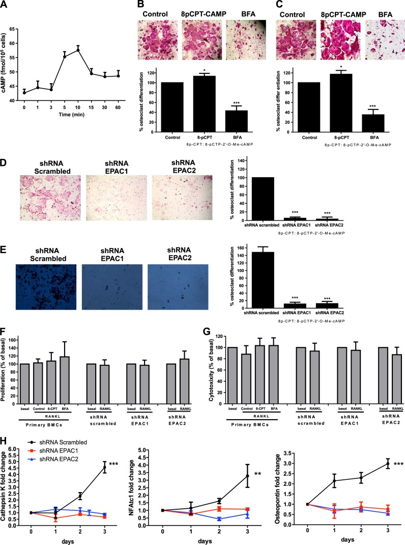
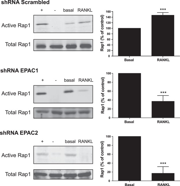
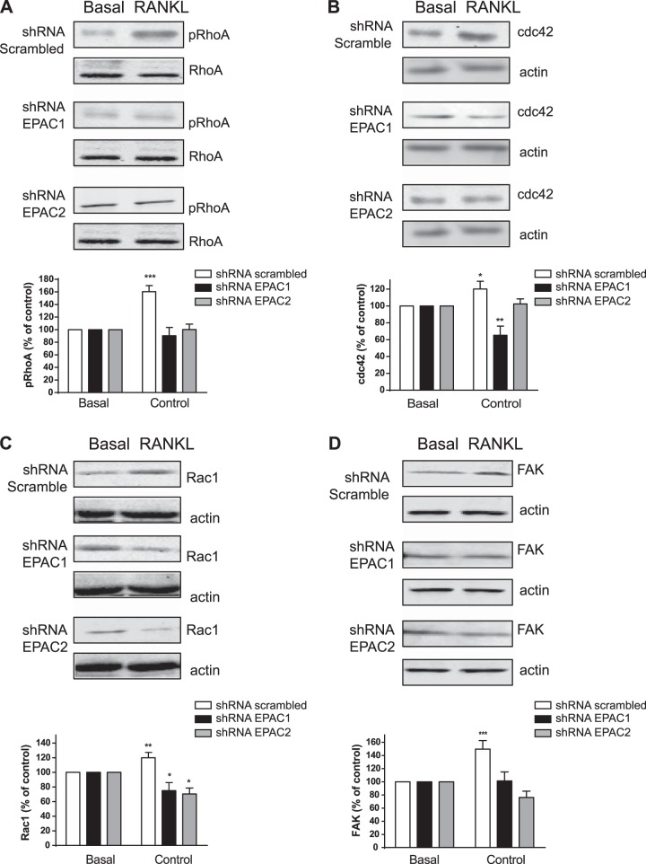
Bisphosphonates inhibit osteoclast differentiation/function via inhibition of Rap1A isoprenylation. As Rap1 is the effector of exchange protein directly activated by cAMP (EPAC) proteins, we determined the role of EPAC in osteoclast differentiation. We examined osteoclast differentiation as the number of primary murine/human bone-marrow precursors that differentiated into multinucleated TRAP-positive cells in the presence of EPAC-selective stimulus (8-pCTP-2'-O-Me-cAMP, 100 μM; 8-pCTP-2'-O-Me-cAMP-AM, 1 μM) or inhibitor brefeldin A (BFA), ESI-05, and ESI-09 (10 μM each). Rap1 activity was assessed, and signaling events, as well as differentiation in EPAC1/2-knockdown RAW264.7 cells, were studied. Direct EPAC1/2 stimulation significantly increased osteoclast differentiation, whereas EPAC1/2 inhibition diminished differentiation (113 ± 6%, P < 0.05, and 42 ± 10%, P < 0.001, of basal, respectively). Rap1 activation was maximal 15 min after RANKL stimulation (147 ± 9% of basal, P < 0.001), whereas silencing of EPAC1/2 diminished activated Rap1 (43 ± 13 and 20 ± 15% of control, P < 0.001) and NFkB nuclear translocation. TRAP-staining revealed no osteoclast differentiation in EPAC1/2-KO cells. Cathepsin K, NFATc1, and osteopontin mRNA expression decreased in EPAC1/2-KO cells when compared to control. RhoA, cdc42, Rac1, and FAK were activated in an EPAC1/2-dependent manner, and there was diminished cytoskeletal assembly in EPAC1/2-KO cells. In summary, EPAC1 and EPAC2 are critical signaling intermediates in osteoclast differentiation that permit RANKL-stimulated NFkB nuclear translocation and actin rearrangements. Targeting this signaling intermediate may diminish bone destruction in inflammatory arthritis.

Keywords: FAK; Rac1; RhoA; cdc42.


Figures

Figure 1.
Figure 1.
EPAC1 and EPAC2 are essential for osteoclast differentiation. A) Cellular cAMP levels over time following stimulation with 50 ng/ml RANKL. cAMP values are expressed as means ± sd (n=6). B) Mouse BMCs treated with 30 ng/ml RANKL together with 8-pCTP-2′-O-Me-cAMP or BFA were fixed and stained for TRAP. TRAP+ cells containing ≥3 nuclei were counted as osteoclasts. C) Primary human BMC-derived osteoclasts were fixed and stained for TRAP following the same conditions as primary cells. D) RAW264.7 cells were stably transduced with scrambled or EPAC1 and EPAC2 shRNA, and the number of RANKL-induced osteoclasts was studied by TRAP staining. E) RAW264.7 cells were stably transduced with scrambled or EPAC1 and EPAC2 shRNA and treated with 50 ng/ml RANKL. Toluidine blue staining was performed to assay osteoclast activity. Results are expressed as mean ± sd number of pits formed. F) Primary cells were challenged with 30 ng/ml RANKL in the presence of 8-pCTP-2′-O-Me-cAMP or BFA, and proliferation was analyzed 48 h later. EPAC1- and EPAC2-knockdown cells were challenged for 48 h with 50 ng/ml RANKL, and proliferation was analyzed. Results are expressed as percentage of basal levels. G) Primary cells were challenged with 30 ng/ml RANKL in the presence of 8-pCTP-2′-O-Me-cAMP or BFA, and cytotoxicity was analyzed 48 h later. EPAC1- and EPAC2- knockdown cells were challenged for 48 h with 50 ng/ml RANKL, and cytotoxicity was analyzed. Results are expressed as percentage of basal levels. H) RAW264.7 cells were stably transduced with scrambled or EPAC1 and EPAC2 shRNA and treated with 50 ng/ml RANKL. Changes in cathepsin K, NFATc1, and osteopontin mRNA during the 3 d osteoclast differentiation process in EPAC1 and EPAC2 shRNA RAW264.7 cells were compared to scrambled shRNA-infected cells. All results are expressed as means ± sd (n=6). *P < 0.05, **P < 0.01, ***P < 0.001 vs. nonstimulated control.
Figure 2.
Figure 2.
RANKL-mediated EPAC activation mediates the expression of the Ras-like small GTPase Rap1. RAW264.7 cells were stably transduced with scrambled or EPAC1 and EPAC2 shRNA and treated with 50 ng/ml RANKL. At 15 min after stimulation, Rap1 pulldown assay was performed, as described in Materials and Methods. Representative Western blots of activated and corresponding total Rap1 protein are shown. Summary data are expressed as mean ± sd percentage of activated Rap1 (percentage of nonstimulated cells; n=4). + indicates positive control; − indicates negative control. **P < 0.01, ***P < 0.001 vs. nonstimulated control.
Figure 3.
Figure 3.
EPAC activation promotes NFκB p50/p105 nuclear translocation. EPAC1, EPAC2, and scrambled silenced RAW264.7 cells were treated with 50 ng/ml of RANKL and p50/p105 NFκB in the cytoplasmic and nuclear cell fractions, IκBα and p-IκBα were studied by Western blot. Results are expressed as means ± sd of 4 independent experiments. To normalize for protein loading, the membranes were reprobed for actin or p84, respectively, and results normalized appropriately. Y axis has been expanded to show the differences more clearly. *P < 0.05, ***P < 0.001 vs. nonstimulated control.
Figure 4.
Figure 4.
Morphological characterization of osteoclast cultures. A) F-actin was detected by Alexa 555-Phalloidin staining. Osteoclasts cultured on glass show 3 distinct morphologies: early stage of least mature osteoclasts (arrow A); an intermediate stage of maturing osteoclasts (arrow B), and the late stage of mature osteoclasts (arrow C). B) RAW264.7 cells stably transduced with EPAC1, EPAC2, or scrambled shRNA were treated with 50 ng/ml of RANKL for 3 d. Cells were plated in fibronectin-coated glass coverslips, and quantitative evaluation of number of least mature, maturing, and mature osteoclasts in osteoclast cultures in EPAC1, EPAC2, and scrambled silenced RAW264.7 cells was performed. Results are expressed as means ± sd of 4 independent experiments. C) F-actin was detected by Alexa 555-phalloidin staining. Images were taken at an original view of ×63. *P < 0.05, **P < 0.01, ***P < 0.001 vs. scrambled cells.
Figure 5.
Figure 5.
EPAC activation promotes RhoA phosphorylation and cdc42 activation. RAW264.7 cells stably transduced with shRNA for EPAC1, EPAC2, or scrambled were treated with 50 ng/ml of RANKL for 15 min. Western blots are representative. A) RhoA phosphorylation was analyzed by Western blot. B) cdc42 activation was studied by Western blot. C) Rac1 activation was analyzed by Western blot. D) FAK activation was studied by Western blot. Results are expressed as mean ± sd of 4 independent experiments. To normalize for protein loading, the membranes were reprobed with actin and results normalized appropriately. *P < 0.05, ***P < 0.001 vs. nonstimulated control.
Figure 6.
Figure 6.
cAMP activation of EPAC promotes osteoclast differentiation by p50/p105 NFκB nuclear translocation and actin rearrangements. Activation of EPAC by the Gs-coupled receptor mediated by RANKL/RANK activation results in activation of p50/p105 NFκB nuclear translocation and the actin cytoskeleton. These events result in promotion of osteoclast differentiation.

References

    1. Suda T., Takahashi N., Udagawa N., Jimi E., Gillespie M. T., Martin T. J. (1999) Modulation of osteoclast differentiation and function by the new members of the tumor necrosis factor receptor and ligand families. Endocr. Rev. 20, 345–357 - PubMed
    1. Manolagas S. C. (2000) Birth and death of bone cells: basic regulatory mechanisms and implications for the pathogenesis and treatment of osteoporosis. Endocr. Rev. 21, 115–137 - PubMed
    1. Teitelbaum S. L. (2000) Bone resorption by osteoclasts. Science 289, 1504–1508 - PubMed
    1. McHugh K. P., Shen Z., Crotti T. N., Flannery M. R., Fajardo R., Bierbaum B. E., Goldring S. R. (2007) Role of cell-matrix interactions in osteoclast differentiation. Adv. Exp. Med. Biol. 602, 107–111 - PubMed
    1. Takayanagi H. (2007) Osteoimmunology: shared mechanisms and crosstalk between the immune and bone systems. Nat. Rev. Immunol. 7, 292–304 - PubMed

Publication types

MeSH terms