

Comment
doi: 10.7554/eLife.03678.
Cryo-EM enters a new era
Affiliations
- PMID: 25122623
- PMCID: PMC4131193
- DOI: 10.7554/eLife.03678
Item in Clipboard
Comment
Cryo-EM enters a new era
Elife.
.
Abstract
Advances in detector hardware and image-processing software have led to a revolution in the use of electron cryo-microscopy to determine complex molecular structures at high resolution.
Keywords: Cryo-EM; image analysis; micoscopy; single-particle analysis.
Copyright © 2014, Kuehlbrandt.
Conflict of interest statement
Figures
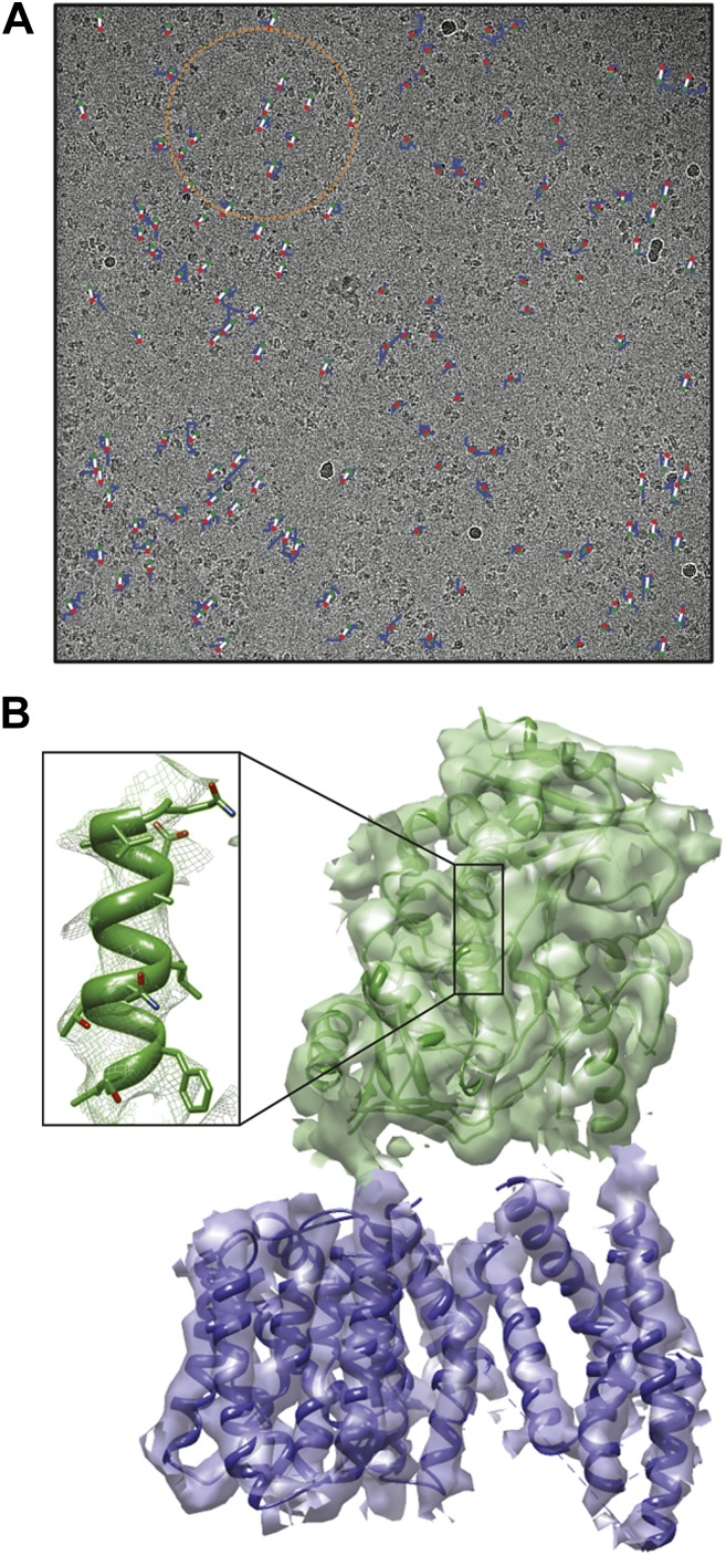
(A) Electron micrograph showing how the γ-secretase particles are moved by the electron beam (blue tracks; movement multiplied by a factor of 50 to make it visible). Scheres and co-workers have developed techniques (Bai et al., 2013; Scheres, 2014) to correct for these movements, and used them to determine the structure of the human γ-secretase complex at a resolution of 4.5 Å (Lu et al., 2014). This approach involves fitting linear tracks to the real movements: the fitted tracks are shown in white, with their start and end points being shown in green and red, respectively; the orange circle outlines the ensemble of particles used for statistical processing to fit the track of one particle (Scheres, 2014). (B) 3D map of the γ-secretase complex. The 19 trans-membrane helices of the four subunits that contain the active site of the complex are shown in blue, and the extracellular domain is shown in green. The inset shows an alpha helix with partly resolved sidechains in the extracellular domain.
Comment on
-
Beam-induced motion correction for sub-megadalton cryo-EM particles.Elife. 2014 Aug 13;3:e03665. doi: 10.7554/eLife.03665. Elife. 2014. PMID: 25122622 Free PMC article.
References
Publication types
MeSH terms
Substances
LinkOut - more resources
Full Text Sources
Other Literature Sources
