Intratumoral injection of Clostridium novyi-NT spores induces antitumor responses
- PMID: 25122639
- PMCID: PMC4399712
- DOI: 10.1126/scitranslmed.3008982
Intratumoral injection of Clostridium novyi-NT spores induces antitumor responses
Abstract
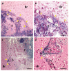
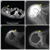
Species of Clostridium bacteria are notable for their ability to lyse tumor cells growing in hypoxic environments. We show that an attenuated strain of Clostridium novyi (C. novyi-NT) induces a microscopically precise, tumor-localized response in a rat orthotopic brain tumor model after intratumoral injection. It is well known, however, that experimental models often do not reliably predict the responses of human patients to therapeutic agents. We therefore used naturally occurring canine tumors as a translational bridge to human trials. Canine tumors are more like those of humans because they occur in animals with heterogeneous genetic backgrounds, are of host origin, and are due to spontaneous rather than engineered mutations. We found that intratumoral injection of C. novyi-NT spores was well tolerated in companion dogs bearing spontaneous solid tumors, with the most common toxicities being the expected symptoms associated with bacterial infections. Objective responses were observed in 6 of 16 dogs (37.5%), with three complete and three partial responses. On the basis of these encouraging results, we treated a human patient who had an advanced leiomyosarcoma with an intratumoral injection of C. novyi-NT spores. This treatment reduced the tumor within and surrounding the bone. Together, these results show that C. novyi-NT can precisely eradicate neoplastic tissues and suggest that further clinical trials of this agent in selected patients are warranted.
Copyright © 2014, American Association for the Advancement of Science.
Figures


Comment in
-
Immunotherapy: the treatment bug--fighting cancer with bacterial infection.Nat Rev Clin Oncol. 2014 Oct;11(10):562. doi: 10.1038/nrclinonc.2014.151. Epub 2014 Sep 2. Nat Rev Clin Oncol. 2014. PMID: 25178634 No abstract available.
-
Therapeutics: Bacterial treatment for cancer.Nat Rev Cancer. 2014 Oct;14(10). doi: 10.1038/nrc3827. Nat Rev Cancer. 2014. PMID: 25230884 No abstract available.
References
-
- Krause DS, Van Etten RA. Tyrosine kinases as targets for cancer therapy. N. Engl. J. Med. 2005;353:172–187. - PubMed
-
- Imai K, Takaoka A. Comparing antibody and small-molecule therapies for cancer. Nat. Rev. Cancer. 2006;6:714–727. - PubMed
-
- Sosman JA, Kim KB, Schuchter L, Gonzalez R, Pavlick AC, Weber JS, McArthur GA, Hutson TE, Moschos SJ, Flaherty KT, Hersey P, Kefford R, Lawrence D, Puzanov I, Lewis KD, Amaravadi RK, Chmielowski B, Lawrence HJ, Shyr Y, Ye F, Li J, Nolop KB, Lee RJ, Joe AK, Ribas A. Survival in BRAF V600–mutant advanced melanoma treated with vemurafenib. N. Engl. J. Med. 2012;366:707–714. - PMC - PubMed
-
- Wilson WR, Hay MP. Targeting hypoxia in cancer therapy. Nat. Rev. Cancer. 2011;11:393–410. - PubMed
-
- Hanahan D, Weinberg RA. Hallmarks of cancer: The next generation. Cell. 2011;144:646–674. - PubMed
Publication types
MeSH terms
Grants and funding
LinkOut - more resources
Full Text Sources
Other Literature Sources
Research Materials

