. 2014 Aug 26;111(34):E3544-52.
doi: 10.1073/pnas.1413201111. Epub 2014 Aug 13.

Calcineurin determines toxic versus beneficial responses to α-synuclein

Affiliations

Calcineurin determines toxic versus beneficial responses to α-synuclein

Gabriela Caraveo et al. Proc Natl Acad Sci U S A. .

Abstract

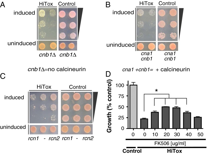
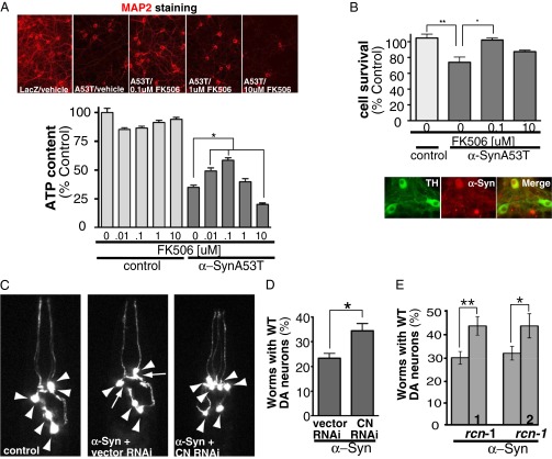
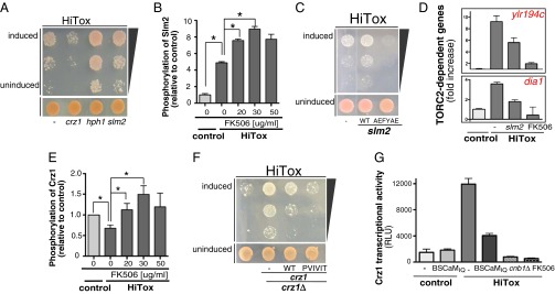
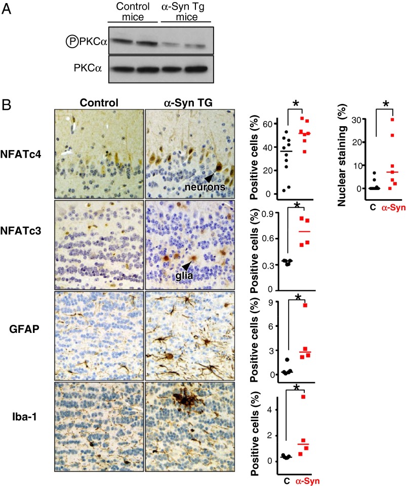
Calcineurin (CN) is a highly conserved Ca(2+)-calmodulin (CaM)-dependent phosphatase that senses Ca(2+) concentrations and transduces that information into cellular responses. Ca(2+) homeostasis is disrupted by α-synuclein (α-syn), a small lipid binding protein whose misfolding and accumulation is a pathological hallmark of several neurodegenerative diseases. We report that α-syn, from yeast to neurons, leads to sustained highly elevated levels of cytoplasmic Ca(2+), thereby activating a CaM-CN cascade that engages substrates that result in toxicity. Surprisingly, complete inhibition of CN also results in toxicity. Limiting the availability of CaM shifts CN's spectrum of substrates toward protective pathways. Modulating CN or CN's substrates with highly selective genetic and pharmacological tools (FK506) does the same. FK506 crosses the blood brain barrier, is well tolerated in humans, and is active in neurons and glia. Thus, a tunable response to CN, which has been conserved for a billion years, can be targeted to rebalance the phosphatase's activities from toxic toward beneficial substrates. These findings have immediate therapeutic implications for synucleinopathies.

Keywords: Crz1; NFAT; Slm2; TORC2; neuroinflammation.


Conflict of interest statement

The authors declare no conflict of interest.

Figures

Fig. 1.
Fig. 1.
BSCaMIQ, a sensor and a sink for calmodulin, rescues toxicity induced by α-syn by decreasing the total levels of free Ca2+–CaM. (A) Strains of control (no α-syn), NoTox (low copy number of α-syn), IntTox (intermediate copy number of α-syn), and HiTox (high copy number of α-syn) were transformed with aequorin, a genetically encoded Ca2+ indicator. Cytosolic Ca2+ was measured by aequorin luminescence over time after α-syn induction. Cytosolic Ca2+ levels are expressed as fold induction relative to control. (B) Cmd1pfree levels assayed by FRET 0, 4, and 8 h after α-syn induction in the presence of BSCaMIQ in control (blue), HiTox strain (red), and HiTox strain transfected with cmd1 (red dashed line). Cmd1pfree = Kd [(Rmax − FRET/CFP)/FRET/CFP − Rmin] (see Supporting Information). (C) Yeast strains were spotted onto plates containing uninducing media [synthetic defined (SD) −Ura; GPD-BSCaMIQ selective; Lower] and replica platted in threefold serial dilutions onto α-syn–inducing plates containing selective media and (SGal −Ura) (Upper). YFP is used as control plasmid. (D) Yeast strains were spotted onto plates containing uninducing media (SD −Ura, Leu; BSCaMIQ and cmd1 selective; Lower) and replica platted in threefold serial dilutions onto α-syn–inducing plates containing selective media and SGal −Ura, Leu (Upper). YFP and empty vector (vec) were used as control plasmids, cmd1 = yeast calmodulin and cmd1X = D94A, E105V; unable to bind calcium at the third EF hand. (E) BSCaMIQ (green), neuronal (red MAP2 positive), and nuclear (blue, Hoechst) stainings from representative pictures of rat primary neuronal cultures coinfected with either control lentivirus LacZ, LacZ and α-SynA53T, LacZ and α-SynA53T and BSCaMIQ, or LacZ and BSCaMIQ. (F) Percentages of neurons (MAP2 positive) relative to control (LacZ infected) in the conditions described in B. *P < 0.05, one-way ANOVA, Dunnett’s multiple comparison test.
Fig. 2.
Fig. 2.
Calcineurin activation is central to α-syn toxicity in yeast. (A) Control and HiTox strains lacking calcineurin (cnb1Δ) were spotted onto plates containing uninducing media (SD −His,Trp–α-syn selective, Lower) and replica plated in threefold serial dilutions on α-syn–inducing plates containing selective media SGal (Upper). (B) Control and HiTox yeast strains were spotted onto plates containing uninducing media (SD −Ura, Leu; cna1- and cnb1 selective; Lower) and replica plated in threefold serial dilutions onto α-syn–inducing plates containing selective media and SGal (Upper). (C) Same assay as in B except control and HiTox strains was plated onto plates containing uninducing media (SD −Leu; rcn1-, or rcn2 selective, respectively). (D) Growth (described as percentage over control) of HiTox cells grown for 48 h over a range of FK506 concentrations. See also Fig. S4D.
Fig. 3.
Fig. 3.
Partial calcineurin activity is necessary to protect against α-syn toxicity ex vivo and in vivo. (A, Upper) MAP2 staining from representative pictures of rat primary neuronal cultures coinfected with a lentivirus carrying LacZ as control and/or α-synA53T treated with various doses of FK506 for 14 d. (Lower) Rat cortical neurons infected with α-synA53T and/or LacZ as control treated with vehicle and/or increasing concentrations of FK506 for 14 d and assayed for viability for ATP content. *P < 0.05, one-way ANOVA, Dunnett’s multiple comparison test. Each data point was normalized against control LacZ at the same FK506 dose. (B, Upper) Dopaminergic neurons from mice infected with α-synA53T and/or mKate as control treated with vehicle and/or distinct concentrations of FK506. Cell survival was measured by counting TH positive neurons after 14 d of α-synA53T infection and drug treatment. *P < 0.05, one-way ANOVA, Dunnett’s multiple comparison test. (Lower) Representative pictures of infection efficiency of mKate α-syn in TH positive neurons. (C) Representative images of C. elegans dopaminergic neurons in worms expressing GFP alone (Left, all six anterior neurons are intact) and in α-syn–expressing worms treated with RNAi control (only four anterior DA neurons remain) or with RNAi for calcineurin (CN) (Right). Arrowheads show intact dopaminergic neuron cell bodies and arrows represent areas where dopaminergic neurons are absent. (D) The population of worms with WT (wild type) numbers of neurons remaining after treatment with RNAi for calcineurin (CN) or empty vector RNAi (control) in α-syn–expressing worms. For each experiment, three independent experiments were performed, with 30 worms per trial. *P < 0.05 (Student t test). See also Fig. S5C. (E) Same assay as in D except with worms overexpressing rcn-1. Bars 1 and 2 represent independent transgenic lines. *P < 0.005, **P < 0.0005 (Student t test). See also Fig. S5B.
Fig. 4.
Fig. 4.
Calcineurin can activate protective and toxic substrates in yeast. (A) The HiTox yeast strain was spotted onto plates containing uninducing media (SD −Leu; vector, hph1, slm2, or crz1 selective; Lower) and replica plated in threefold serial dilutions onto α-syn–inducing plates containing selective media and SGal −Leu (Upper). (B) Peak intensity of phosphopeptide ENVD(phospho)SPR from Slm2p relative to control using SRM-mass spectrometry after phosphopeptide enrichment as described (66). Error bars reflect biological and technical variability. *P < 0.05 (Student t test). (C) Similar spotting assays as described in A but onto plates containing uninducing media [SD −Leu; slm2, slm2-AEFYAE (unable to bind calcineurin) selective; Lower] and replica plated in threefold serial dilutions onto α-syn–inducing plates containing selective media and SGal −Leu (Upper). (D) Real-time PCR for the TORC2-dependent genes ylr194c and dia1 in control and HiTox yeast strains in the presence of slm2 or a low dose of FK506 (25 μg/mL). (E) Peak intensity of phosphopeptide MDSANS(phospho)SEKISK from Crz1p relative to control using SRM-mass spectrometry after phosphopeptide enrichment as described (66). Error bars reflect technical variability. *P < 0.05 (Student t test). (F) The HiTox yeast strain was spotted onto plates containing uninducing media [SD −Leu; vector, crz1, PVIVIT-crz1 (high affinity to calcineurin) selective; Lower] and replica plated in threefold serial dilutions onto α-syn–inducing plates containing selective media and SGal −Leu (Upper) in the presence and absence of crz1 (crz1Δ). (G) β-Glactosidase luminescent assay for control and HiTox strains harboring a reporter for crz1p-dependent transcriptional activity in the presence and absence of 25 μg/mL FK506, calcineurin (cnb1Δ), and/or BSCaMIQ.
Fig. 5.
Fig. 5.
Calcineurin substrates in α-syn transgenic mice. (A) Western blot for phosphoserine 657, PKCα from brain lysates of 12-mo-old α-syn transgenic mice. PKCα serves as loading control. (B) Immunohistochemistry for NFATc4 in neurons from the mitral cell layer in the olfactory bulb of 13-mo-old α-syn transgenic mice and control (C) mice (Upper). Immunohistochemistry for NFATc3 in glia from the internal plexiform layer in the olfactory bulb of 13-mo-old α-syn transgenic. Immunohistochemistry for glial fibrillary acid protein (GFAP) for normal and reactive astrocytes in the internal plexiform layer of the olfactory bulb. Similarly, immunostaining for Iba-1 from normal and activated microglia in the nodules of the internal plexiform layer of the olfactory bulb of α-syn transgenic mice. Error bars reflect variability between two sections analyzed from a total of five animals in each group. *P < 0.05 (Student t test).
Fig. 6.
Fig. 6.
NFATc4 nuclear expression is increased in cases of PD and DLB. Immunohistochemistry for NFATc4 staining in neurons from the substantia nigra pars compacta, hippocampus area CA3, and layers 5 and 6 of the frontal cortex in human PD and DLB cases. An average of two sections from five control (C) and eight diseased cases were analyzed. Nuclear staining was scored by a neuropathologist. Scoring: 0, no nuclear staining; 1, scattered positive nuclei; 2, positive nuclear <30% of neurons; 3, positive nuclei focally >30% of neurons. n, neuronal nucleus; nm, neuromelanin; nuc, nucleolus. (Scale bar, 50 μm.) *P < 0.05 (Student t test).

References

    1. Wojda U, Salinska E, Kuznicki J. Calcium ions in neuronal degeneration. IUBMB Life. 2008;60(9):575–590. - PubMed
    1. Adamczyk A, Strosznajder JB. Alpha-synuclein potentiates Ca2+ influx through voltage-dependent Ca2+ channels. Neuroreport. 2006;17(18):1883–1886. - PubMed
    1. Danzer KM, et al. Different species of alpha-synuclein oligomers induce calcium influx and seeding. J Neurosci. 2007;27(34):9220–9232. - PMC - PubMed
    1. Martin ZS, et al. α-Synuclein oligomers oppose long-term potentiation and impair memory through a calcineurin-dependent mechanism: Relevance to human synucleopathic diseases. J Neurochem. 2012;120(3):440–452. - PMC - PubMed
    1. Buttner S, et al. The Ca2+/Mn2+ ion-pump PMR1 links elevation of cytosolic Ca2+ levels to alpha-synuclein toxicity in Parkinson's disease models. Cell Death Differ. 2012;20(3):465–77. - PMC - PubMed

Publication types

MeSH terms

LinkOut - more resources