. 2014 Sep 30;111(39):14147-52.
doi: 10.1073/pnas.1404171111. Epub 2014 Aug 13.

Arginine starvation-associated atypical cellular death involves mitochondrial dysfunction, nuclear DNA leakage, and chromatin autophagy

Affiliations

Arginine starvation-associated atypical cellular death involves mitochondrial dysfunction, nuclear DNA leakage, and chromatin autophagy

Chun A Changou et al. Proc Natl Acad Sci U S A. .

Abstract

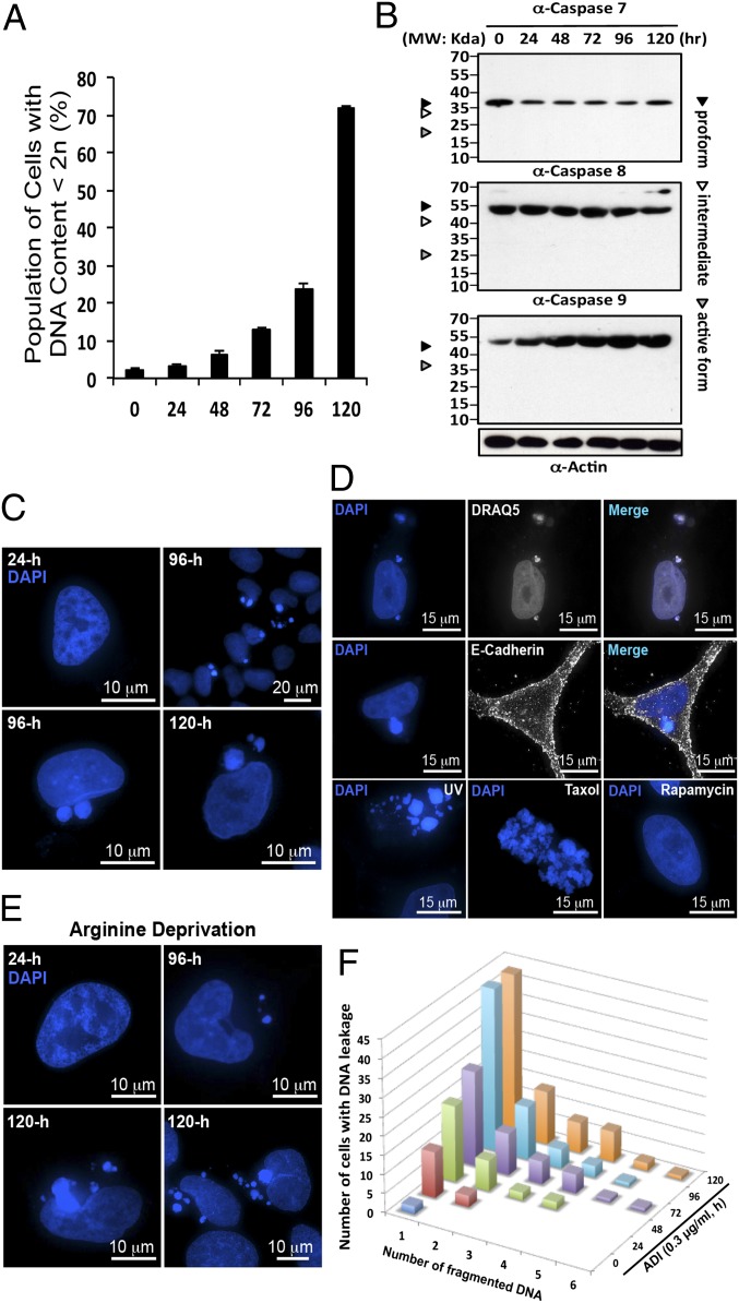
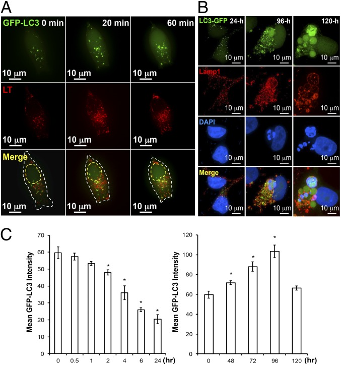
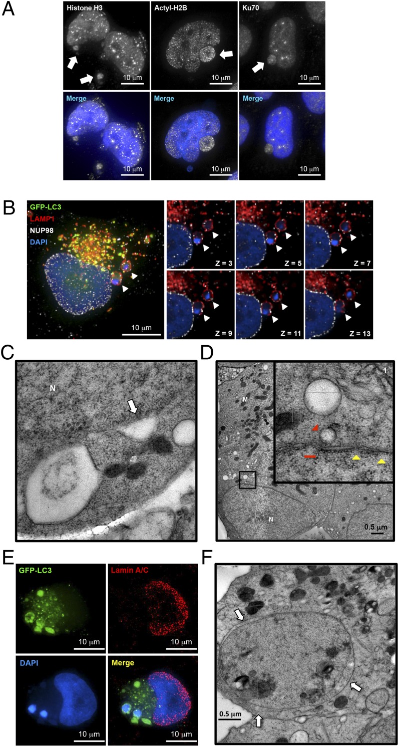
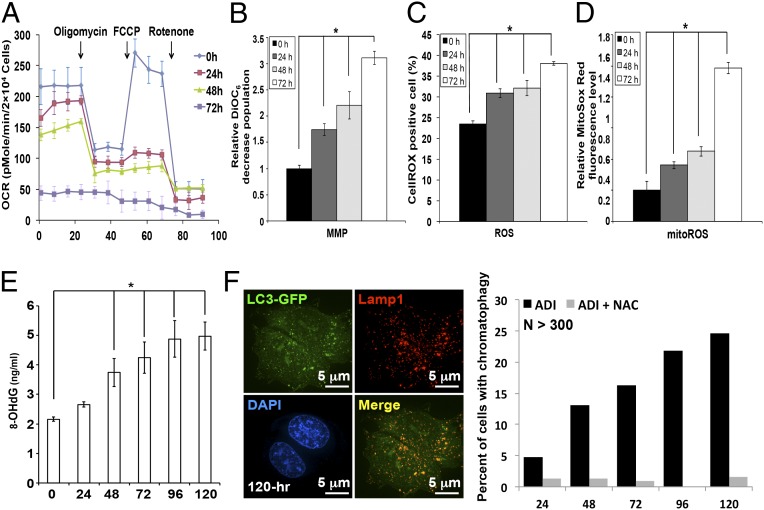
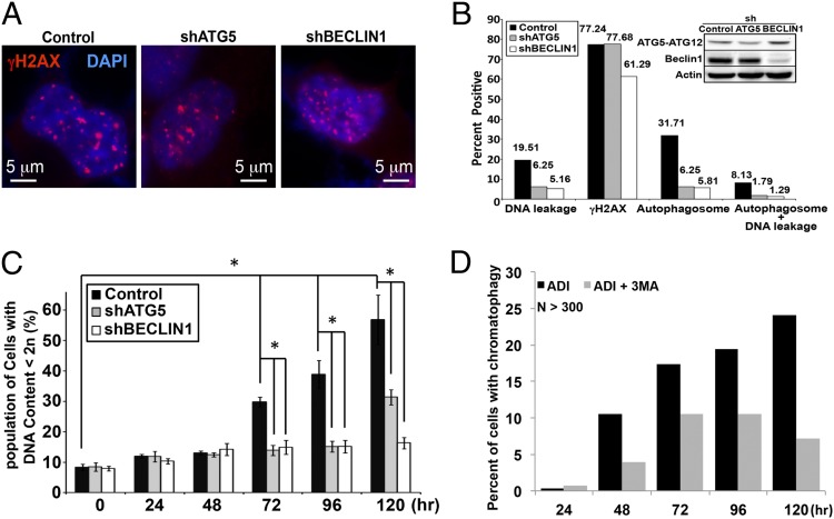
Autophagy is the principal catabolic prosurvival pathway during nutritional starvation. However, excessive autophagy could be cytotoxic, contributing to cell death, but its mechanism remains elusive. Arginine starvation has emerged as a potential therapy for several types of cancers, owing to their tumor-selective deficiency of the arginine metabolism. We demonstrated here that arginine depletion by arginine deiminase induces a cytotoxic autophagy in argininosuccinate synthetase (ASS1)-deficient prostate cancer cells. Advanced microscopic analyses of arginine-deprived dying cells revealed a novel phenotype with giant autophagosome formation, nucleus membrane rupture, and histone-associated DNA leakage encaptured by autophagosomes, which we shall refer to as chromatin autophagy, or chromatophagy. In addition, nuclear inner membrane (lamin A/C) underwent localized rearrangement and outer membrane (NUP98) partially fused with autophagosome membrane. Further analysis showed that prolonged arginine depletion impaired mitochondrial oxidative phosphorylation function and depolarized mitochondrial membrane potential. Thus, reactive oxygen species (ROS) production significantly increased in both cytosolic and mitochondrial fractions, presumably leading to DNA damage accumulation. Addition of ROS scavenger N-acetyl cysteine or knockdown of ATG5 or BECLIN1 attenuated the chromatophagy phenotype. Our data uncover an atypical autophagy-related death pathway and suggest that mitochondrial damage is central to linking arginine starvation and chromatophagy in two distinct cellular compartments.

Keywords: ADI-PEG20; arginine auxotrophy; cancer therapy; metabolic stress; prostate cancer.


Conflict of interest statement

The authors declare no conflict of interest.

Figures

Fig. 1.
Fig. 1.
ADI-PEG20 induced both caspase-independent cell death and DNA leakage. (A) CWR22Rv1 cells were treated with ADI-PEG20 and stained with propidium iodide (PI). The percentage of hypodiploid cells began to rise at 48 h and significantly increased after 72 h. (B) Western blotting showing the lack of caspase 7, 8, and 9 activation in cells treated with ADI-PEG20. (C) DAPI staining reveals nuclear DNA before leakage at 24 h (early time points) and after leakage at 96 h and 120 h (late time points) posttreatment. (D) The same samples were stained with another fluorescent DNA dye, DRAQ5. Cells were stained with plasma membrane marker E-cadherin to reveal its boundary. Cells were treated with UV or Taxol (1 nM) to demonstrate the appearance of apoptotic body and mitotic catastrophe, respectively. Rapamycin (2 μM) treatment did not produce the same effect. (E) Prolong incubation with arginine-depleted medium also induced the same phenotype. (F) The bar graph illustrates the positive increment of leaked DNA particles in a population of cells showing the phenotype.
Fig. 2.
Fig. 2.
Prolonged ADI-PEG20 treatment can induce giant-autophagosome formation and affect autophagic flux pattern. (A) Representative time-lapse images showing ADI-PEG20 treatment induces autophagy and facilitates the fusion of autophagosome (green) and lysosome (red, LysoTracker) into autophagolysosome (yellow). The outline is shown by a dashed white line. The estimated nucleus location is shown by a dashed yellow line. (B) Prolonged ADI-PEG20 treatment induces abnormally sized autophagosomes (green), which colocalize with lysosome (red) and leaked DNA (blue). (C) ADI-PEG20 treatment induces autophagic flux with distinct kinetics. Mean GFP-LC3 intensity in each group was plotted as a bar graph. Data were collected from three independent experiments and are shown as mean ± SD; *P < 0.05.
Fig. 3.
Fig. 3.
Prolonged arginine deprivation by ADI-PEG20 induces nuclear membrane remodeling. (A) Immunofluorescence analysis showed that histone H3, acetylated H2B, and Ku70 were found associated with the exo-nucleus DNA cluster. (B) Immunofluorescence analysis of NUP98 (white), GFP-LC3 (green), lysosome (red), and DAPI (blue) showed that uniform NUP98 signals were both around and outside of the nucleus and colocalized with lysosome signal (white arrowheads). (C) TEM demonstrated that autophagosomes could potentially fuse with the nuclear membrane (white arrow). (D) Autophagosomes were found in close proximity to the nucleus (red arrowhead, with higher magnification shown in inset). Nucleus membrane also showed partial breakages (red line, ∼150 nm). A typical nuclear pore complex has a thin layer of electron dense disk with an opening about 120 nm; however, it may appear smaller under Cryo-EM (35) (yellow arrowheads, 47 nm ∼ 53 nm). M, mitochondria; N, nucleus. (E) Representative immunofluorescence images showed reduced lamin signals at the site of excessive autophagy. (F) Abnormal nucleus membrane morphology was found in treated cells at later time points. Expansion of intramembrous space was observed between inner membrane and outer membrane (white arrows). One representative cyro-EM image is shown in C, D, and F. n = 3 in AF.
Fig. 4.
Fig. 4.
ADI-PEG20 treatment impairs mitochondrial function and induces ROS production. (A) ADI-PEG20 treatment suppresses basal OCR and reserve capacity. The tracing revealed the basal OCR decreased in a time-dependent manner with the incubation of ADI-PEG20. (BD) ADI-PEG20 impairs mitochondrial membrane potential (MMP) and elevates ROS. Respective MMP (B) CellROX oxidation (C), and MitoSox Red oxidation (D) were determined by flow cytometry after staining with DiOC6, DCFDA, and MitoSOX Red. Data are collected from three independent experiments and shown as mean ± SD; *P < 0.05. (E) The increase of 8-OHdG after ADI-PEG20 treatment. Data were collected from three independent experiments with mean ± SD; *P < 0.05. (F) ROS scavenger NAC attenuated DNA leakage phenotype (Right; n > 300) and formation of giant autophagosomes (Left).
Fig. 5.
Fig. 5.
Autophagy contributes to ADI-induced DNA leakage. (A) Control, shATG5, and shBECLIN1 stably transfected CWR22Rv1 cells overexpressing GFP-LC3 (green) were treated with ADI-PEG20 for 96 h, followed by staining with anti-γH2AX antibody (red) and DAPI (blue). (B) Approximately 120–150 cells were analyzed for DNA leakage, γH2AX staining, and autophagosome formation. Western blot of ATG5-ATG12 and BECLIN1 demonstrates the expression of these proteins in modified cells. (C) PI staining revealed that knocking down autophagy machinery significantly reduces hypodiploid cell population after 72 h. (D) The bar graph shows the interruption of autophagolysosome formation with 3-MA diminished the DNA-leakage phenotype (n > 250).

Comment in

References

    1. Kim JW, Dang CV. Cancer’s molecular sweet tooth and the Warburg effect. Cancer Res. 2006;66(18):8927–8930. - PubMed
    1. Vander Heiden MG, Cantley LC, Thompson CB. Understanding the Warburg effect: The metabolic requirements of cell proliferation. Science. 2009;324(5930):1029–1033. - PMC - PubMed
    1. Dang CV. Rethinking the Warburg effect with Myc micromanaging glutamine metabolism. Cancer Res. 2010;70(3):859–862. - PMC - PubMed
    1. Feun L, et al. Arginine deprivation as a targeted therapy for cancer. Curr Pharm Des. 2008;14(11):1049–1057. - PMC - PubMed
    1. Huang CC, et al. Arginine deprivation as a new treatment strategy for head and neck cancer. Oral Oncol. 2012;48(12):1227–1235. - PubMed

Publication types

MeSH terms