. 2014;42(16):10809-22.
doi: 10.1093/nar/gku745. Epub 2014 Aug 13.

Molecular insights into replication initiation by Qβ replicase using ribosomal protein S1

Affiliations

Molecular insights into replication initiation by Qβ replicase using ribosomal protein S1

Daijiro Takeshita et al. Nucleic Acids Res. 2014.

Abstract

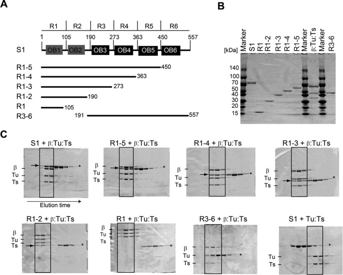
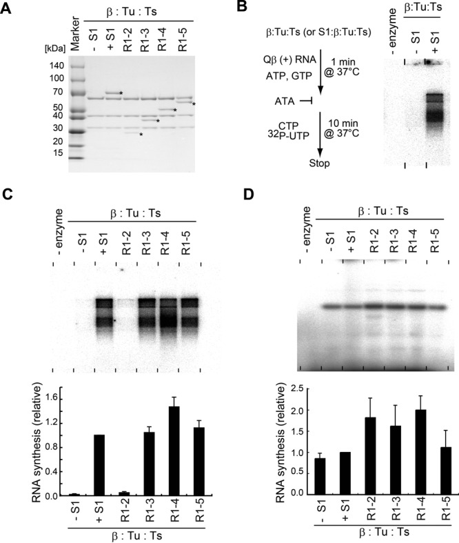
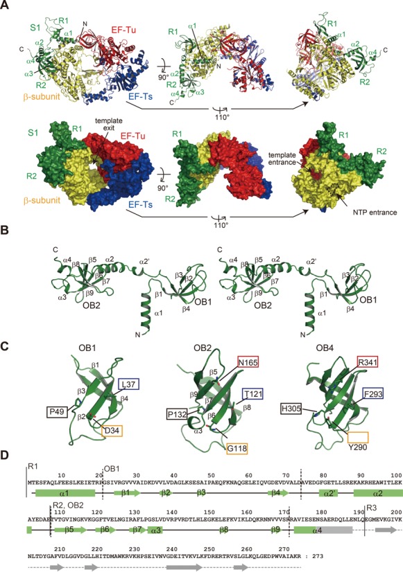
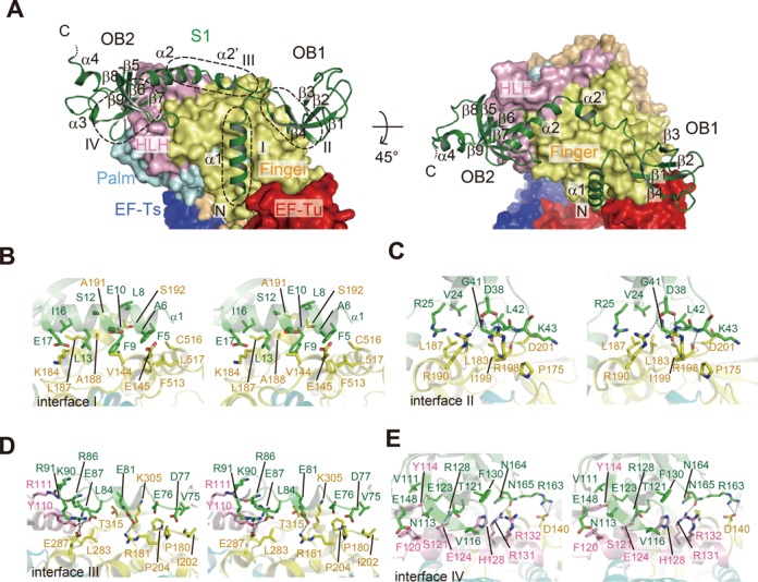
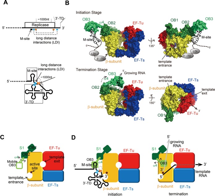
Ribosomal protein S1, consisting of six contiguous OB-folds, is the largest ribosomal protein and is essential for translation initiation in Escherichia coli. S1 is also one of the three essential host-derived subunits of Qβ replicase, together with EF-Tu and EF-Ts, for Qβ RNA replication in E. coli. We analyzed the crystal structure of Qβ replicase, consisting of the virus-encoded RNA-dependent RNA polymerase (β-subunit), EF-Tu, EF-Ts and the N-terminal half of S1, which is capable of initiating Qβ RNA replication. Structural and biochemical studies revealed that the two N-terminal OB-folds of S1 anchor S1 onto the β-subunit, and the third OB-fold is mobile and protrudes beyond the surface of the β-subunit. The third OB-fold mainly interacts with a specific RNA fragment derived from the internal region of Qβ RNA, and its RNA-binding ability is required for replication initiation of Qβ RNA. Thus, the third mobile OB-fold of S1, which is spatially anchored near the surface of the β-subunit, primarily recruits the Qβ RNA toward the β-subunit, leading to the specific and efficient replication initiation of Qβ RNA, and S1 functions as a replication initiation factor, beyond its established function in protein synthesis.


Figures

Figure 1.
Figure 1.
Interaction of S1 with the core Qβ replicase. (A) Schematic representation of ribosomal protein S1, consisting of six OB-fold motifs, and its variants used for the present study. (B) Purification of the S1 protein, its variants and the core Qβ replicase (β:Tu:Ts). Proteins were resolved by 4–20% (v/v) SDS polyacrylamide gel electrophoresis (PAGE) and stained with Coomassie Brilliant Blue (CBB). (C) Analysis of the interaction of core Qβ replicase with S1 variants by size-exclusion chromatography. The fractions were separated by 10% (v/v) or 12% (v/v) SDS-PAGE and stained with CBB. The arrows in the solid square in the gels indicate S1 or its variants bound to the core Qβ replicase (β:Tu:Ts) complex. Asterisks (*) in the gels indicate the bands of S1 or its variants.
Figure 2.
Figure 2.
Minimal domains of S1 for Qβ negative RNA synthesis. (A) Purification of reconstituted complexes of core Qβ replicase and S1 variants. Protein complexes were resolved by 4–20% (v/v) SDS PAGE and stained with CBB. Asterisks (*) indicate the band of S1 and its variants. (B) In vitro S1-dependent negative strand RNA synthesis. Simplified flowchart of the assay. ATA, aurintricarboxylic acid, is an inhibitor of RNA synthesis initiation by Qβ replicase
Figure 3.
Figure 3.
Complex structure of the core Qβ replicase and the N-terminal half of S1. (A) Overall structure of QβSN, the β-subunit, EF-Tu, EF-Ts and the N-terminal domains (R1 and R2) of S1. R3 of the S1 protein was not clearly visible in the structure. Ribbon models (upper panel) and surface models (lower panel). (B) Stereo view of the structure of R1–3. R3 was not visible in the determined structure. (C) Comparison of the structures of OB1 (left), OB2 (middle) and OB4 (right, 16, PDB ID: 2KHI). The amino acid residues putatively involved in RNA binding by OB4 are depicted, and the amino acid residues located at the corresponding positions in OB1 and OB2 are also shown. (D) Sequence of R1–3 of S1 along with the secondary structures. α-helices and β-sheets in R1 and R2 are depicted by solid squares and arrows, respectively. The secondary structure of the R3 domain was predicted based on the sequence alignments.
Figure 4.
Figure 4.
Interactions between the N-terminal half of S1 and the β-subunit. (A) Structure of R1–3 bound to the β-subunit, depicted by aribbon model. The finger, helix-loop-helix (HLH) and palm domains of the β-subunit are depicted by surface models. Contact interfaces between R1–3 of S1 and the β-subunit are depicted by dotted circles (interfaces I–IV). (B)(E) Stereo views of the detailed interactions between the β-subunit and the S1 protein at each interface in (A).
Figure 5.
Figure 5.
The N-terminal half of S1 binds the internal region of Qβ RNA. (A) Schematic presentation of the gene organization of Qβ RNA (18). Secondary structure of the M-site RNA used in the present study. The numbering of the RNA is that of the positive Qβ RNA. (B) In vitro negative strand RNA synthesis from positive strand Qβ RNA, in the presence of various amounts of M-site RNA (left) or tRNA (right). Reaction mixtures were separated as in Figure 2B. The graph below indicates the quantification of 32P-labeled RNA products in the gels. (C) The M-site RNA gel-retardation assays of full-length S1 and its variants in Figure 1A. (D) UV cross-linking of the M-site RNA and Qβ replicase containing S1 variants. The graph below indicates the quantification of 32P-labeled S1 or S1 variants. The bars in the graphs in (B) and (D) are the standard deviations of more than two independent experiments.
Figure 6.
Figure 6.
RNA-binding activity of OB3 for negative strand Qβ RNA synthesis. (A) A structural model of the OB-fold in R3 (upper). The amino acid residues putatively involved in RNA binding are shown in stick models. Structure of the S1 domain of ribonuclease II complexed with RNA (42). Residues involved in RNA binding are depicted by sticks. (B) The M-site RNA gel-retardation assays by the R1–3 domains of S1 and its mutants. The graph indicates the quantifications of M-site RNA fractions bound to R3 and its mutants. (C)In vitro negative strand synthesis by complexes of core Qβ replicase and R1–3 of S1 and its variants. Reaction mixtures were separated as in Figure 2B. (D) In vitro RNA synthesis using DN3 RNA as the template RNA, as in Figure 2D. The bars in the graphs in (C) and (D) are the standard deviations of more than two independent experiments.
Figure 7.
Figure 7.
Mechanism of initiation of negative strand Qβ RNA synthesis. (A) One-dimensional simplified view of the 3′-half of Qβ RNA (above) and two-dimensional view of the 3′-half of Qβ RNA. The long distance interactions (LDI) in the RNA (18,19) are depicted by dotted lines, and the regions used for interactions in the Qβ RNA are shown. 3′-TD stands for 3′-terminal domain. (B) Structure of Qβ replicase containing R1–3. The mobile R3 was modeled. The R3 is capable of pivotal rotation, using the α helix (α4) between OB2 and OB3 as a swing arm for both initiation and termination of stages. The template Qβ RNA at the replication initiation stage (upper) and the template and growing RNAs at the replication termination stage (lower) are modeled. LDI and 3′-TD in Qβ RNA were simplified. See the Discussion section in the text. (C) Simplified cartoon of Qβ replicase containing the N-terminal half of S1. The β-subunit, EF-Tu, EF-Ts and S1 (OB1 and OB2) and the mobile OB3 of S1 are depicted. (D) A simplified model of the initiation of negative strand RNA synthesis (left). R3 interacts with the M-site of the Qβ RNA. A simplified model of RNA synthesis termination (right). The interaction between the growing RNA and the mobile R3 of S1 at the termination stage triggers the release of the RNA product from the complex (43).

Similar articles

Cited by

References

    1. Subramanian A.R. Structure and functions of ribosomal protein S1. Prog. Nucleic Acid Res. Mol. Biol. 1983;28:102–138. - PubMed
    1. Farwell M.A., Robert M.W., Rabinowitz J.C. The effect of ribosomal protein S1 from Escherichia coli and Micrococcus luteus on protein synthesis in vitro by E. coli and Bacillus subtilis. Mol. Microbiol. 1992;6:3375–3383. - PubMed
    1. Boni I.V., Isaeva D.M., Musychenko M.L., Tzareva N.V. Ribosome-messenger recognition: mRNA target sites for ribosomal protein S1. Nucleic Acids Res. 1991;19:155–162. - PMC - PubMed
    1. Tedin K., Resch A., Blasi U. Requirements for ribosomal protein S1 for translation initiation of mRNAs with and without a 5′ leader sequence. Mol. Microbiol. 1997;25:189–199. - PubMed
    1. Sorensen M.A., Fricke J., Pedersen S. Ribosomal protein S1 is required for translation of most, if not all, natural mRNAs in Escherichia coli in vivo. J. Mol. Biol. 1998;280:561–569. - PubMed

Publication types

Associated data

LinkOut - more resources