. 2014 Aug 13;34(33):10884-91.
doi: 10.1523/JNEUROSCI.4795-13.2014.

High-NaCl perception in Drosophila melanogaster

Affiliations

High-NaCl perception in Drosophila melanogaster

Georges Alves et al. J Neurosci. .

Abstract

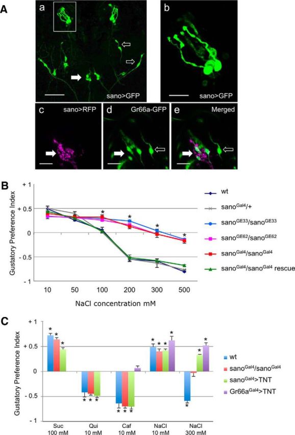
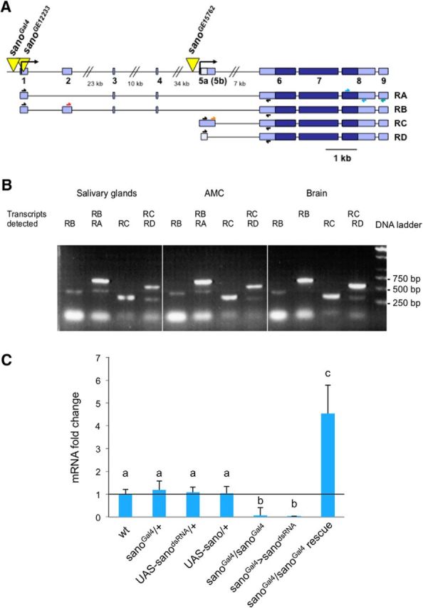
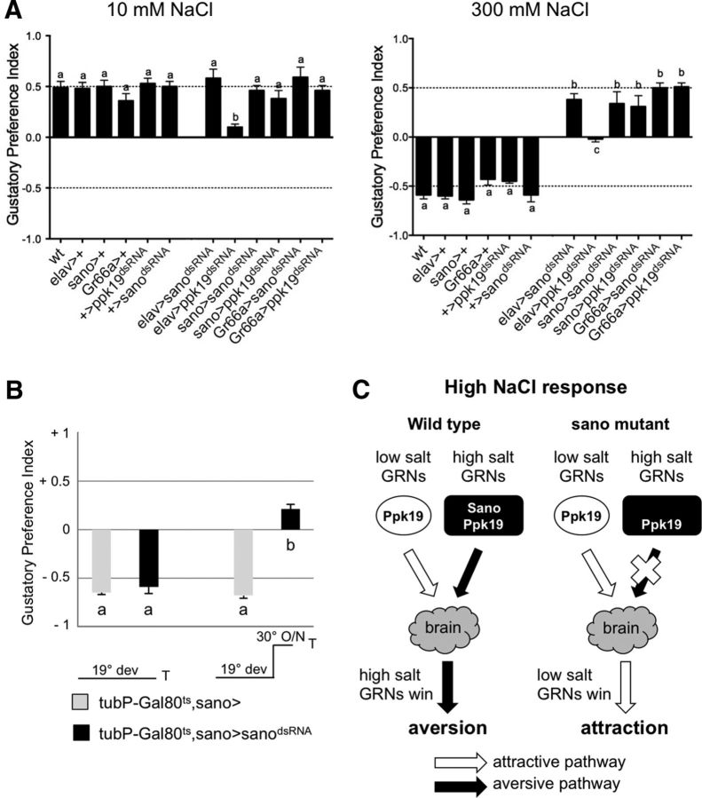
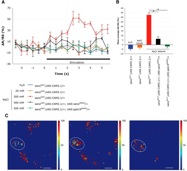
Salt is a fundamental nutrient that is required for many physiological processes, including electrolyte homeostasis and neuronal activity. In mammals and Drosophila, the detection of NaCl induces two different behaviors: low-salt concentrations provide an attractive stimulus, whereas high-salt concentrations are avoided. We identified the gene called serrano (sano) as being expressed in the sensory organs of Drosophila larvae. A transgenic reporter line showed that sano was coexpressed with Gr66a in a subset of gustatory neurons in the terminal organ of third-instar larvae. The disruption of sano gene expression in gustatory neurons led to the specific loss of high-salt concentration avoidance in larvae, whereas the detection of other attractive or aversive substances was unaffected. Moreover, using a cellular marker sensitive to calcium levels, Sano function was shown to be required for neuronal activity in response to high-salt concentrations. In these neurons, the loss of the DEG/ENaC channel PPK19 function also eliminated the cellular response to high-salt concentrations. Our study revealed that PPK19 and Sano are required in the neurons of the larval gustatory organs for the detection of high-salt concentrations.

Keywords: Drosophila melanogaster; behavior; chemosensory system; larva; salt; taste.


Figures

Figure 1.
Figure 1.
Role of sano in taste perception. Aa, Ab, Ventral view of the anterior part of a third-instar sanoGal4/sanoGal4;10XUAS–IVS–mCD8::GFP/10XUAS–IVS-mCD8::GFP larva. Aa, Expression is observed in gustatory neurons: five neurons in the TO (box) and three neurons in the ventral pharyngeal sense organ (filled arrow). Expression is also present in few neurons along the body (open arrows). Scale bar, 50 μm. Ab, Detailed view of the expression in five neurons of the TO. Scale bar, 10 μm. Ac–Ae, Ventral view of the anterior part of a third-instar sanoGal4/+;10XUAS–IVS–mCD8::RFP/Gr66a–GFP larva. Ac, sanoGal4 drives expression of RFP in six neurons of the TO ganglion. Ad, Gr66a–GFP drives expression of GFP in four neurons of the TO ganglion (filled arrow) and in two adjacent neurons of the TO dorsolateral group (open arrow). Ae, sanoGal4 and Gr66a–GFP are coexpressed in four neurons of the TO ganglion (filled arrow). B, Behavioral responses of third-instar larvae to NaCl concentrations. The GPIs for indicated NaCl concentrations versus water were calculated. For each condition, 10 trials with 50 larvae were performed. Control lines (wild-type; sanoGal4/+), sano mutants (sanoGal4/sanoGal4; sanoGE12233/sanoGE12233; sanoGE15762/sanoGE15762), and the sano rescue line (sanoGal4/sanoGal4;UAS–sano/+) were used in this experiment. Each bar represents a mean ± SEM of GPI (n = 10). Asterisks above error bars indicate that results were significantly different (ANOVA, p < 0.05). C, Behavioral responses of third-instar larvae to attractive [100 mm sucrose (Suc) or 10 mm NaCl] or repulsive [10 mm caffeine (Caf), 10 mm quinine (Qui), or 300 mm NaCl] tastants. Control larvae (wild-type), sano mutant larvae (sanoGal4/sanoGal4), and larvae with impaired gustatory neurons (sanoGal4>TNT or Gr66aGal4>TNT) were tested. Each point represents the mean ± SEM of GPI obtained from 10 trials of 50 larvae. A Student's t test comparing with the theoretical value 0 was performed. Asterisks above error bars indicate that results were significantly different (p < 0.05).
Figure 2.
Figure 2.
Map and transcript analysis of sano gene. A, Structure of the sano gene. Yellow triangles represent the P-element insertions [sanoGal4 is a P{GawB} line, and sanoGE12233 and sanoGE15762 are P{UAS} lines]. Exons are shown in blue. Light blue boxes represent the 5′ and 3′ untranslated regions. Scale bar, 1 kb. The arrows above the transcripts indicate the primers used for the RT-PCR analysis. B, sano expression analyzed by RT-PCR. RNAs were extracted from the dissected structures of the CantonS larvae. A PCR with a primer specific to the sano sequence was performed to obtain cDNAs by reverse transcription. All of the four transcripts were present in the different organs tested. AMC, Antenno-maxillary complex. C, The data represent the fold change in the amount of sano transcripts determined by qPCR. The CantonS strain was used as the wild-type reference, and the level of wild-type transcripts was defined as 1.0. The error bars represent the SEM, n = 3–10. The same letter over error bars indicates values that are not significantly different (ANOVA, p < 0.05).
Figure 3.
Figure 3.
Disruption of sano and ppk19 by dsRNA. A, Behavioral assay for 10 mm NaCl and 300 mm NaCl. The effects of sano or ppk19 disruption in all larval neurons (elavGal4 crosses), sano-expressing neurons (sanoGal4 crosses), or Gr66a-expressing neurons (Gr66aGal4 crosses) were tested in these experiments. Each histogram (mean ± SEM of GPI) was calculated from 10 trials. The same letter over error bars indicates values that were not significantly different (ANOVA, p < 0.05). B, The effects of the temporal disruption of Sano on aversive behavior at 300 mm NaCl. Two temperature conditions were used. The offspring from the crosses were raised at 19°C (19° dev). At 19°C, GAL4 activity is repressed by GAL80ts such that sano transcripts are not impaired in tubP–Gal80ts,sano>sanodsRNA larvae. When tubP–Gal80ts,sano>sanodsRNA larvae are transferred to a permissive temperature of 30°C overnight (O/N), sanoGal4 drives expression of sanodsRNA. Each histogram (mean ± SEM of GPI) was calculated from 10 trials. The same letter over the error bars indicates values that were not significantly different (ANOVA, p < 0.05). C, Schematic model showing that the loss of Sano selectively impairs the aversive salt taste pathway. ppk19 is required in both low- and high-salt GRNs to detect NaCl. sano is required in high-salt GRNs to detect high-NaCl concentrations. In wild-type context, both low- and high-salt GRNs are activated by high-NaCl concentration, leading to aversion. Loss of sano selectively disrupts the aversive pathway, leading to attraction in the presence of high-NaCl concentration.
Figure 4.
Figure 4.
Sano and PPK19 are required for TO neuron activation by high-NaCl concentrations. A, Representative fluorescence change (ΔR/R0) of CAM2.1 expressed in sanoGal4 neurons in control and mutant larvae. Neuronal activity was measured before and after the addition of different stimuli: distilled water, 20 mm NaCl, or 300 mm NaCl. Responses to 300 mm NaCl were measured in larvae expressing either sanodsRNA or ppk19dsRNA in sanoGal4 neurons (n = 4–6). The error bars represent the SEM. B, Mean FRET ratio changes. Changes were measured during the first 5 s of the stimulation of different control and mutant larvae (n = 4–6). The error bars represent the SEM. Asterisks indicate that results are significantly different (Student's t test, p < 0.05). C, Peak responses are shown in false-color scale (ΔR/R0%, right of images). Left, sanoGAL4,UAS–CAM2.1/+ before stimulation. Middle, sanoGAL4,UAS–CAM2.1/+ at 2 s after stimulation with 300 mm NaCl. Right, sanoGAL4,UAS–CAM2.1/+;UAS--sanodsRNA/+ at 2 s after stimulation with 300 mm NaCl. The cell bodies of the neurons used for the FRET measurements are circled. Scale bar, 10 μm.

References

    1. Amrein H, Thorne N. Gustatory perception and behavior in Drosophila melanogaster. Curr Biol. 2005;15:R673–R684. doi: 10.1016/j.sbi.2005.10.006. - DOI - PubMed
    1. Balakireva M, Gendre N, Stocker RF, Ferveur JF. The genetic variant Voila causes gustatory defects during Drosophila development. J Neurosci. 2000;20:3425–3433. - PMC - PubMed
    1. Chandrashekar J, Kuhn C, Oka Y, Yarmolinsky DA, Hummler E, Ryba NJ, Zuker CS. The cells and peripheral representation of sodium taste in mice. Nature. 2010;464:297–301. doi: 10.1038/nature08783. - DOI - PMC - PubMed
    1. Chung S, Vining MS, Bradley PL, Chan CC, Wharton KA, Jr, Andrew DJ. Serrano (sano) functions with the planar cell polarity genes to control tracheal tube length. PLoS Genet. 2009;5:e1000746. doi: 10.1371/journal.pgen.1000746. - DOI - PMC - PubMed
    1. Colomb J, Grillenzoni N, Ramaekers A, Stocker RF. Architecture of the primary taste center of Drosophila melanogaster larvae. J Comp Neurol. 2007;502:834–847. doi: 10.1002/cne.21312. - DOI - PubMed

Publication types

LinkOut - more resources