. 2014 Aug 13;34(33):11159-72.
doi: 10.1523/JNEUROSCI.0180-14.2014.

Amyloid precursor protein dimerization and synaptogenic function depend on copper binding to the growth factor-like domain

Affiliations

Amyloid precursor protein dimerization and synaptogenic function depend on copper binding to the growth factor-like domain

Frederik Baumkötter et al. J Neurosci. .

Abstract

Accumulating evidence suggests that the copper-binding amyloid precursor protein (APP) has an essential synaptic function. APP synaptogenic function depends on trans-directed dimerization of the extracellular E1 domain encompassing a growth factor-like domain (GFLD) and a copper-binding domain (CuBD). Here we report the 1.75 Å crystal structure of the GFLD in complex with a copper ion bound with high affinity to an extended hairpin loop at the dimerization interface. In coimmunoprecipitation assays copper binding promotes APP interaction, whereas mutations in the copper-binding sites of either the GFLD or CuBD result in a drastic reduction in APP cis-orientated dimerization. We show that copper is essential and sufficient to induce trans-directed dimerization of purified APP. Furthermore, a mixed culture assay of primary neurons with HEK293 cells expressing different APP mutants revealed that APP potently promotes synaptogenesis depending on copper binding to the GFLD. Together, these findings demonstrate that copper binding to the GFLD of APP is required for APP cis-/trans-directed dimerization and APP synaptogenic function. Thus, neuronal activity or disease-associated changes in copper homeostasis likely go along with altered APP synaptic function.

Keywords: Alzheimer's disease; amyloid precursor protein; copper; dimerization; metal homeostasis; synaptogenesis.


Figures

Figure 1.
Figure 1.
Copper binding to the GFLD. A, Crystal structure of the GFLD in complex with copper in a color ramp from the N terminus (amino acid 28; blue) to the C terminus (amino acid 189; red). The N-terminal 6×His tag is not included in the diagram representation. The anomalous difference map for the copper ion (11.3 σ) and coordinating histidine residues H108 and H110 within the LBL (yellow-orange) are shown. B, Electrostatic surface potential (3 kT) around the copper-binding site (arrow); hydrophobic (gray) and positively (blue) or negatively (red) charged surface patches are indicated. C, Superposition of the LBL in the apo (gray; PDB identification number 1mwp) and copper-bound (color ramp) forms. Note the copper-induced reduced state of C98 and C105 as indicated by the final 2Fo-Fc map shown at 1.0 σ, the disintegration of the β-sheets, and the insertion of two stabilizing water molecules (magenta). D, Thermal unfolding monitored by CD spectroscopy. The E1 domain without (blue) or saturated with (red) copper ions was heated from 20°C to 90°C, and the ellipticity (θ) at 222 nm was measured. Differences in the unfolding process between copper-bound and apo E1 indicate that copper influences the fully reversible thermal unfolding of the E1 domain. E, Sequence alignment of the LBL from selected APP family members (hs, human; cp, guinea pig; mm, mouse; ce, Caenorhabditis elegans; dm, Drosophila melanogaster). The N-terminal and C-terminal numbering of each LBL sequence is listed. Conserved cysteine residues forming the disulfide bond in the LBL are colored in yellow, and the copper-binding H×H motif is highlighted in green. F, The copper coordination sphere. The copper ion is fivefold coordinated in a square pyramid formed by H108 and H110, as well as D23 of a symmetry-related molecule.
Figure 2.
Figure 2.
Copper binds to the GFLD of APP. A, Scheme of the APP subdomains used for ITC. The positions of the GFLD (light gray), CuBD (dark gray), and the N-terminal GST epitope (black circle) are indicated. The protease cleavage site is indicated by an arrow, and the sites of mutation are indicated by an asterisk. B, SDS-PAGE of the affinity-purified APP subdomains stained with Coomassie blue. C–H, ITC data recorded at 25°C. Titration of glycine-complexed CuCl2 into 20 μm APP E1 (C), 13 μm APP CuBD (D), 14 μm APP GFLD (E), HEPES buffer as control (F), 38 μm APP GFLD H108/110A (GFLDmut) (G), or 43 μm APP E1 H108/110/147/151A (APP E1mut) (H). The panels in C (left) and D and E (top) show the differential heating power (Δp) versus time (t) plot. C (right), D,E (bottom), Normalized heat of reaction (Q) versus molar copper/protein ratio (R). Copper binds with high affinities to the APP E1 domain (KD = 7.5 nm), the CuBD (KD = 18 nm), and the GFLD (KD = 28 nm). Binding to the APP GFLD or APP E1 is completely abolished by substitution of the copper-coordinating histidines H108/H110 or H108/H110/H147/H151 with alanine residues, respectively.
Figure 3.
Figure 3.
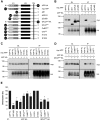
Copper promotes APP dimerization. A, Analysis of cell viability monitored by LDH release. HEK293 cells were incubated for 4 h in growth medium supplemented with the indicated amounts of copper, and LDH released into the medium was measured. Maximal LDH released into the medium was determined after cell lysis with Triton X-100 (+) (n = 3). B, Cellular copper uptake monitored by ICP-OES. HEK293 cells were incubated for 4 h in growth medium supplemented with the indicated amounts of copper, and the cellular copper concentration normalized to protein levels was determined (n = 3). C, Co-IP analysis of HA- and myc-tagged APP with increasing copper concentrations. HEK293 cells were transiently cotransfected with C-terminal HA-tagged (APP HA) and N-terminal myc-tagged (myc APP) APP and incubated with the indicated amounts of glycine-complexed CuCl2 for 4 h. Anti-HA immunoprecipitates from cell extracts (IP) were immunoblotted for HA- and myc-tagged proteins (DL, direct load). D, Quantification of Co-IP analysis revealed significantly increased dimerization in the presence of 250 μm copper. Mean protein levels were normalized to APP ± SEM (n ≥ 3; unpaired Student's t test, **p < 0.01). E, F, Co-IP analyses of HA- and myc-tagged APP with increasing concentrations of zinc (E) and iron (II) (F).
Figure 4.
Figure 4.
Mutations of copper-binding sites do not affect cellular trafficking of APP. A, Scheme of APP constructs used. ΔE1, APP lacking the E1 domain; Δ91-111, APP lacking the LBL; GFLDmut, APP H108/110A; CuBDmut, APP H147/151A; C/Gmut, APP H108/110A and H147/151A; E1, E1 domain (gray); E2, E2 domain (black); TM, transmembrane domain; *, site of mutation; myc, N-terminal fused c-myc epitope. The positions of the GFLD (light gray) and CuBD (dark gray) are indicated. B, Western blot analysis of APP constructs expressed in HeLa cells. APP proteins were detected by W0-2 antibody and actin by AC-15 antibody. C, Fluorescence micrographs of HeLa cells expressing the different myc-tagged APP constructs (green). Heterologously expressed APP was visualized by an anti-myc antibody and the ER by coexpression of mRFP-tagged KDEL fusion protein (red). D, Pearson's correlation coefficient of APP with mRFP–KDEL. Bars represent mean ± SEM values of three independent experiments (ANOVA with Tukey's HSD post hoc test, **p < 0.01).
Figure 5.
Figure 5.
Intracellular APP dimerization depends on copper binding. Co-IP analysis of HA- and myc-tagged APP in HEK293 cells. A, Scheme of APP constructs used (as described in Fig. 4). APP HA, HA-tagged full-length APP; APP FL, myc-tagged full-length APP; ΔGFLD, APP lacking the GFLD; ΔCuBD, APP lacking the CuBD; HA, C-terminal fused HA epitope; myc, N-terminal fused c-myc epitope. The positions of the GFLD (light gray) and CuBD (dark gray) are indicated. HA-tagged full-length APP (APP HA) was transiently coexpressed with APP FL, ΔE1, ΔGFLD, or ΔCuBD (B) and APP FL, GFLDmut, CuBDmut, C/Gmut, APP H137A, or APP H183A (C). D, Mutated HA-tagged APP (GFLDmut HA) was transiently coexpressed with APP FL, GFLDmut, CuBDmut, or C/Gmut. Anti-HA immunoprecipitates from cell extracts (IP) were immunoblotted for HA- and myc-tagged proteins. DL, direct load. E, Quantification of Co-IP analysis revealed significant changes between APP FL and all APP constructs with mutations/deletions of the copper-binding sites. Mean protein levels were normalized to APP FL ± SEM (n ≥ 4; unpaired Student's t test, *p < 0.05, **p < 0.01).
Figure 6.
Figure 6.
Copper promotes APP trans-dimerization. A, Scheme of APPex fused to the FC domain of human IgG1 (FC) (APPex-FC). B, Coomassie blue-stained SDS-PAGE of the APPex-FC (arrowhead) purification from conditioned medium of COS7 cells (COS7 SN) stably expressing APPex-FC by protein A and heparin affinity chromatography. FT, Flow through; E, eluate; *, MW of IgG heavy chain. C, Representative micrographs of clustered protein A beads coated with either FC or APPex-FC in the presence or absence of 100 μm copper or 2 μm heparin dodecasaccharide (dp12). Scale bar, 10 μm. D, Quantification of clustered protein A beads incubated with the indicated amounts of copper, iron, or zinc, as well as Heparin dp12 or EDTA. Aggregates >10 μm were measured with a Coulter Counter Z2 particle counter. Aperture tube, 100 μm. Bars represent mean ± SEM values of at least three independent experiments (unpaired Student's t test, *p < 0.05 and **p < 0.01 compared with APPex-FC-coated beads in HEPES buffer; ##p < 0.01 compared with APPex-FC-coated beads in HEPES buffer with 100 μm CuCl2). E, GPC of purified APP GFLD (black) and APP GFLD H108/110A (GFLDmut; gray) in the absence (solid line) or presence of either 10-fold molar excess of heparin dodecasaccharide (dp12; dashed line) or 50-fold molar excess of CuCl2 (broken line). The median apparent MWs and the elution volumes of the marker proteins carbonic anhydrase (Mr = 29,000) and ribonuclease A (Mr = 13,700) are indicated (▾).
Figure 7.
Figure 7.
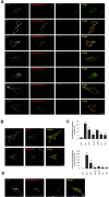
Mutation of APP copper-binding sites decreases APP synaptogenic activity. Hemisynapse analysis of APP synaptogenic activity. A, Representative micrographs of immunostainings of HEK293 cells expressing either GFP, Neuroligin-1 (Nlg), APP FL, APP lacking the E1 domain (ΔE1), APP lacking the entire extracellular domain (ΔEC), APP H108/110A (GFLDmut), or APP H147/151A (CuBDmut), cocultured with primary cortical neurons. Cells were stained with anti-Synaptophysin (red) and anti-MAP2 (blue) antibodies. Synaptophysin-positive but MAP2-negative puncta are visible on top of Nlg- and APP-transfected cells (green). Scale bar, 10 μm. B, Immunostaining of APP-expressing HEK293 cells cocultured with primary cortical neurons. Cells were stained with anti-SV2 (red) and anti-Synapsin-1 (red) antibodies to demonstrate the recruitment of presynaptic marker proteins. Scale bar, 10 μm. C, Quantification of Synaptophysin-positive puncta per transfected HEK293 cell and Synaptophysin-covered area per HEK293 cell (5-7 cells per experiment). Bars represent mean ± SEM values of at least three independent experiments (ANOVA with Tukey's HSD post hoc test, *p < 0.05, **p < 0.01, and ***p < 0.001 compared with GFP; #p < 0.05 and ##p < 0.01 compared with APP FL). D, APP-expressing HEK293 cells cocultured with primary cortical neurons were stained with anti-Synaptophysin (red) and anti-Tau1 (blue) antibodies to visualize an accumulation of Synaptophysin at contact sites between the axon and HEK293 cells. Scale bar, 10 μm.
Figure 8.
Figure 8.
Model for copper-induced APP dimerization at the synapse. A, Under resting conditions, the copper transporter ATP7A is predominantly localized at the Golgi apparatus, and the copper concentration within the synaptic cleft is low (light blue). Under these conditions, APP present at presynaptic and postsynaptic sites is mainly monomeric. B, NMDA receptor activation during synaptic transmission causes an ATP7A-mediated release of copper into the synaptic cleft (dark blue). Our results suggest that increasing copper concentrations promote dimerization of APP in both cis- and trans-cellular manner contributing to APP synaptogenic activity. In the insets, the domain organization of monomeric and dimeric APP is shown. The N-terminal E1 domain, subdivided in the GFLD (gray) and the CuBD (black), is linked via a flexible acidic region to the E2 domain, the juxtamembrane/TM region, and the cytosolic domain. The copper-binding sites in the GFLD and CuBD (indicated by blue circles) are crucial for cis- and trans-interactions. The TM region could additionally contribute to lateral dimerization and the E2 domain to trans-cellular dimerization. CTR1, Copper transporter 1; NMDAR, NMDA receptor; PM, plasma membrane.

Similar articles

Cited by

References

    1. Adams PD, Afonine PV, Bunkóczi G, Chen VB, Davis IW, Echols N, Headd JJ, Hung LW, Kapral GJ, Grosse-Kunstleve RW, McCoy AJ, Moriarty NW, Oeffner R, Read RJ, Richardson DC, Richardson JS, Terwilliger TC, Zwart PH. PHENIX: a comprehensive Python-based system for macromolecular structure solution. Acta Crystallogr D Biol Crystallogr. 2010;66:213–221. doi: 10.1107/S0907444909052925. - DOI - PMC - PubMed
    1. Altan-Bonnet N, Sougrat R, Liu W, Snapp EL, Ward T, Lippincott-Schwartz J. Golgi inheritance in mammalian cells is mediated through endoplasmic reticulum export activities. Mol Biol Cell. 2006;17:990–1005. doi: 10.1091/mbc.E05-02-0155. - DOI - PMC - PubMed
    1. Banci L, Bertini I, Ciofi-Baffoni S, Kozyreva T, Zovo K, Palumaa P. Affinity gradients drive copper to cellular destinations. Nature. 2010;465:645–648. doi: 10.1038/nature09018. - DOI - PubMed
    1. Battye TG, Kontogiannis L, Johnson O, Powell HR, Leslie AG. iMOSFLM: a new graphical interface for diffraction-image processing with MOSFLM. Acta Crystallogr D Biol Crystallogr. 2011;67:271–281. doi: 10.1107/S0907444910048675. - DOI - PMC - PubMed
    1. Baumgartner W, Hinterdorfer P, Ness W, Raab A, Vestweber D, Schindler H, Drenckhahn D. Cadherin interaction probed by atomic force microscopy. Proc Natl Acad Sci U S A. 2000;97:4005–4010. doi: 10.1073/pnas.070052697. - DOI - PMC - PubMed

Publication types

Associated data

LinkOut - more resources