. 2014;42(16):10823-31.
doi: 10.1093/nar/gku756. Epub 2014 Aug 14.

Why double-stranded RNA resists condensation

Affiliations

Why double-stranded RNA resists condensation

Igor S Tolokh et al. Nucleic Acids Res. 2014.

Abstract

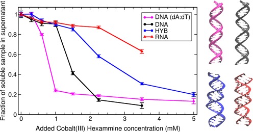
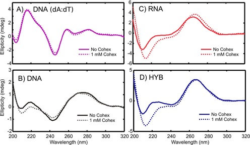
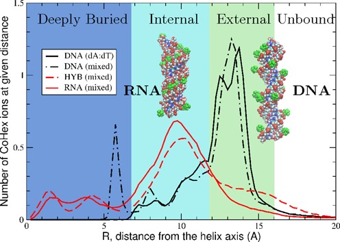
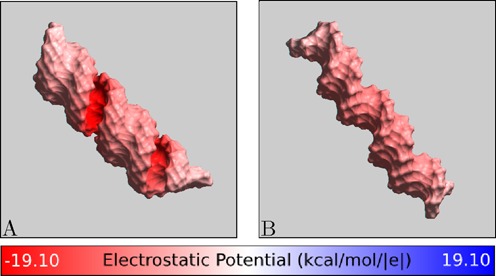
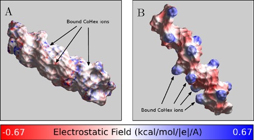
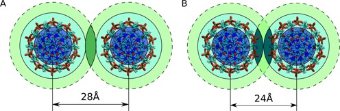
The addition of small amounts of multivalent cations to solutions containing double-stranded DNA leads to inter-DNA attraction and eventual condensation. Surprisingly, the condensation is suppressed in double-stranded RNA, which carries the same negative charge as DNA, but assumes a different double helical form. Here, we combine experiment and atomistic simulations to propose a mechanism that explains the variations in condensation of short (25 base-pairs) nucleic acid (NA) duplexes, from B-like form of homopolymeric DNA, to mixed sequence DNA, to DNA:RNA hybrid, to A-like RNA. Circular dichroism measurements suggest that duplex helical geometry is not the fundamental property that ultimately determines the observed differences in condensation. Instead, these differences are governed by the spatial variation of cobalt hexammine (CoHex) binding to NA. There are two major NA-CoHex binding modes--internal and external--distinguished by the proximity of bound CoHex to the helical axis. We find a significant difference, up to 5-fold, in the fraction of ions bound to the external surfaces of the different NA constructs studied. NA condensation propensity is determined by the fraction of CoHex ions in the external binding mode.


Figures

Figure 1.
Figure 1.
Fraction of unprecipitated short 25 bp NA duplexes (DNA homopolymer, mixed sequence DNA, RNA and DNA:RNA hybrid) calculated from UV absorption as a function of CoHex concentration in solution (starting duplex concentration is 40 μM). The condensation propensity of each duplex is characterized by the CoHex concentration at the midpoint of duplex condensation—the higher the value the lower the propensity. The right panel illustrates the overall suggested structures of these duplexes as either B-form or A-like form helices. The color matches the lines in the panel on the left.
Figure 2.
Figure 2.
CD spectra of the NA helices with and without CoHex.
Figure 3.
Figure 3.
CoHex distributions and ion binding modes (shells) around four types of NA duplexes. DNA duplexes exhibit mostly external CoHex binding (12–16 Å). For RNA and DNA:RNA hybrid, most neutralizing CoHex ions are bound internally (7–12 Å). Shown are the average numbers of CoHex ions in thin (0.25 Å) cylindrical layers around duplexes, at the given distance R from the helical axis. Insets show representative snapshots of RNA (left) and DNA (right) structures with bound CoHex ions (green). In DNA, 80–100% of bound ions are localized at the surface of the phosphate backbone.
Figure 4.
Figure 4.
Electrostatic potential at the ‘CoHex-accessible’ surface of (A) A-form RNA and (B) B-form DNA structures, without bound CoHex ions. Shown is the electrostatic potential computed 3 Å away (CoHex radius) from the molecular surface. B-DNA minor groove is sterically inaccessible to large CoHex ions.
Figure 5.
Figure 5.
Charge neutralization patterns of NA duplexes by bound CoHex ions, assessed by the strength of the electric field near the NA-CoHex complex surface. (A) A-form mixed sequence RNA with CoHex counterions, which bind mostly in the major groove. (B) B-form mixed sequence DNA with CoHex ions, which are bound mostly externally. The specific snapshots are chosen to illustrate the internal (A) and external (B) binding modes from Figure 3 and reflect the actual average binding preferences; each snapshot has 15 bound (near neutralizing) CoHex ions, and is taken from the corresponding 320 ns-long all-atom MD simulation described in ‘Materials and Methods’. See Supplementary Data for a detailed visual characterization of CoHex ion distributions around these structures. The field is computed 3 Å away from the NA-CoHex complex molecular surface.
Figure 6.
Figure 6.
Schematic of (A) the external–external and (B) external–internal CoHex shell overlaps at different interaxial distances. The shell colors correspond to Figure 3. The overlapping shell regions are indicated by a darker color to guide the eye to the differences between (A) and (B).

References

    1. Bloomfield V.A. Condensation of DNA by multivalent cations: considerations on mechanism. Biopolymers. 1991;31:1471–1481. - PubMed
    1. Bloomfield V.A. DNA condensation by multivalent cations. Biopolymers. 1997;44:269–282. - PubMed
    1. Wong G.C.L., Pollack L. Electrostatics of strongly charged biological polymers: ion-mediated interactions and self-organization in nucleic acids and proteins. Ann. Rev. Phys. Chem. 2010;61:171–189. - PubMed
    1. Luger K., Mader A.W., Richmond R.K., Sargent D.F., Richmond T.J. Crystal structure of the nucleosome core particle at 2.8 Å resolution. Nature. 1997;389:251–260. - PubMed
    1. Belyi V.A., Muthukumar M. Electrostatic origin of the genome packing in viruses. Proc. Natl. Acad. Sci. 2006;103:17174–17178. - PMC - PubMed

Publication types

MeSH terms