. 2014 Oct 3;289(40):27952-65.
doi: 10.1074/jbc.M114.581199. Epub 2014 Aug 14.

Biosynthesis and translocation of unsulfated acyltrehaloses in Mycobacterium tuberculosis

Affiliations

Biosynthesis and translocation of unsulfated acyltrehaloses in Mycobacterium tuberculosis

Juan Manuel Belardinelli et al. J Biol Chem. .

Abstract

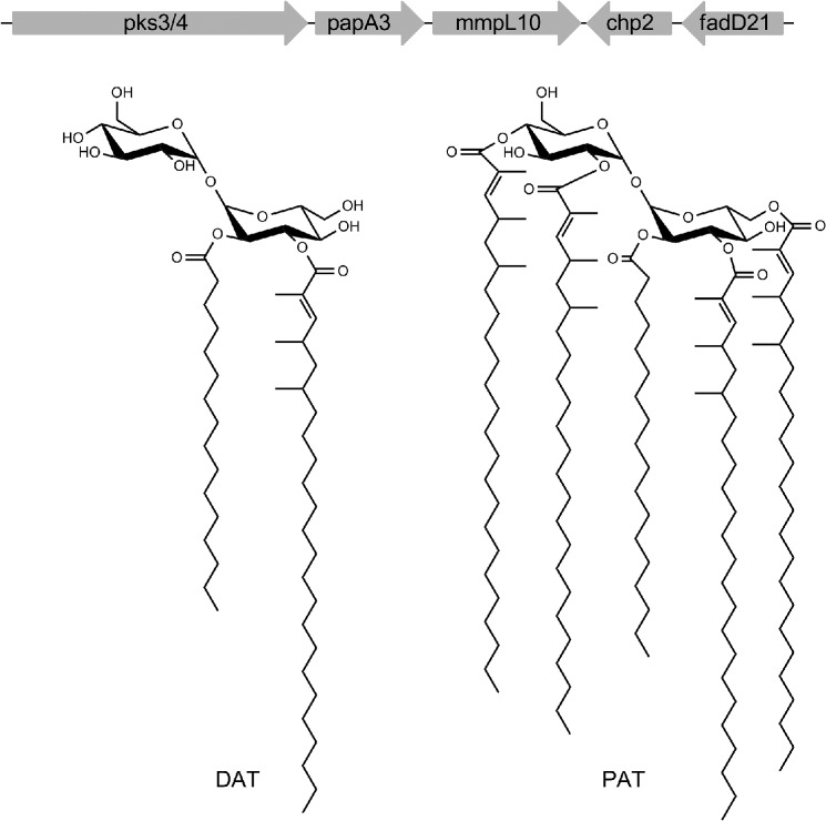
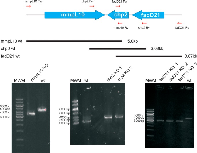
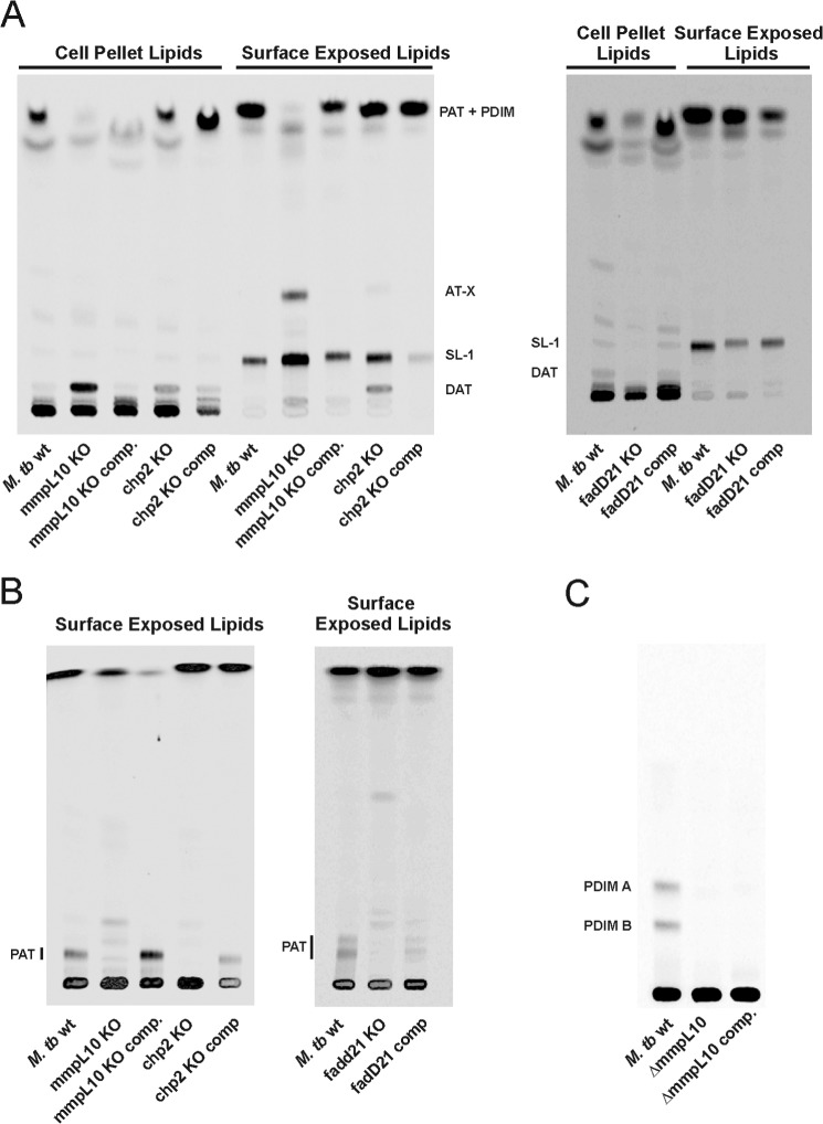
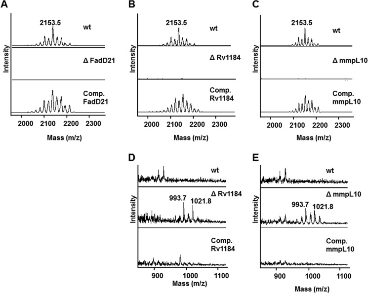
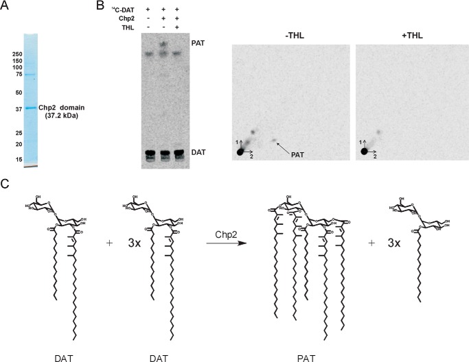
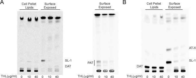
A number of species-specific polymethyl-branched fatty acid-containing trehalose esters populate the outer membrane of Mycobacterium tuberculosis. Among them, 2,3-diacyltrehaloses (DAT) and penta-acyltrehaloses (PAT) not only play a structural role in the cell envelope but also contribute to the ability of M. tuberculosis to multiply and persist in the infected host, promoting the intracellular survival of the bacterium and modulating host immune responses. The nature of the machinery, topology, and sequential order of the reactions leading to the biosynthesis, assembly, and export of these complex glycolipids to the cell surface are the object of the present study. Our genetic and biochemical evidence corroborates a model wherein the biosynthesis and translocation of DAT and PAT to the periplasmic space are coupled and topologically split across the plasma membrane. The formation of DAT occurs on the cytosolic face of the plasma membrane through the action of PapA3, FadD21, and Pks3/4; that of PAT occurs on the periplasmic face via transesterification reactions between DAT substrates catalyzed by the acyltransferase Chp2 (Rv1184c). The integral membrane transporter MmpL10 is essential for DAT to reach the cell surface, and its presence in the membrane is required for Chp2 to be active. Disruption of mmpL10 or chp2 leads to an important build-up of DAT inside the cells and to the formation of a novel form of unsulfated acyltrehalose esterified with polymethyl-branched fatty acids normally found in sulfolipids that is translocated to the cell surface.

Keywords: Acyltransferase; Acyltrehalose; Chp2; Glycolipid; MmpL10; Mycobacteria; Polyketide; Transporter; Tuberculosis.


Figures

FIGURE 1.
FIGURE 1.
DAT and PAT structures and biosynthetic gene cluster. Genes associated with DAT and PAT biosynthesis and export are clustered on the M. tuberculosis H37Rv chromosome. In the forms of DAT and PAT represented here, trehalose is esterified with palmitic acid and multimethyl-branched mycolipenic acids.
FIGURE 2.
FIGURE 2.
Disruption of the fadD21, chp2 (Rv1184c), and mmpL10 genes of M. tuberculosis H37Rv mc26206. 1–3 candidate mutants obtained for each of the three genes were analyzed by PCR. The expected sizes of the PCR fragments for the wild-type parent strain are indicated in the schematic representation of the DAT/PAT locus. A 1.2-kb kanamycin-resistance cassette replaces the ORFs in each of the knock-out mutants. Thus, sizes are 3.87 kb for the wild-type parent strain and 3.12 kb for the knock-out mutants in the case of fadD21; 3.06 kb for the wild-type parent and 3.44 kb for the knock-out mutants in the case of chp2; and 5.0 kb for the wild-type parent and 3.5 kb for the knock-out mutants in the case of mmpL10. MWM, molecular weight marker. wt, wild-type parent strain.
FIGURE 3.
FIGURE 3.
Effects of knocking out fadD21, chp2, and mmpL10 on DAT and PAT biosynthesis and export in M. tuberculosis. Thin layer chromatograms of surface-exposed and cell pellet-associated lipids derived from [1-14C]propionate-labeled wild-type, mutant, and complemented mutant strains. 10,000 cpm were loaded per lane. TLC plates were developed in CHCl3/CH3OH/H2O (90:10:1, v/v/v) for DAT, SL-1, and AT-X analysis (A) or in petroleum ether/acetone (92:8, v/v) for PAT analysis and revealed by phosphorimaging (B). C, thin layer chromatogram of total lipids derived from [1-14C]propionate-labeled wild-type, mmpL10 mutant, and complemented mmpL10 mutant strains showing the absence of PDIM in the mutant and complemented mutant strains. The TLC plate was developed in petroleum ether/diethyl ether (95:5, v/v).
FIGURE 4.
FIGURE 4.
MALDI-MS analysis of lipids extracted from the wild-type, knock-out mutant, and complemented mutant strains. Total lipids extracted from wild-type M. tuberculosis H37Rv mc26206; the fadD21, chp2 (Rv1184), and mmpL10 knock-out mutants; and the complemented mutant strains were precipitated with acetone and subjected to MALDI-MS analysis in the positive ion mode as described under “Experimental Procedures.” Peaks observed are m/z 2153.5, corresponding to sodium-cationized PAT, and m/z 993.7 and 1021.8, corresponding to sodium-cationized DAT. DAT accumulate in the cell pellet-associated lipids of the mmpL10 and chp2 mutant strains as well as in the surface-exposed lipids of the chp2 knock-out mutant. No DAT or PAT were detected in the fadD21 mutant. A–C correspond to surface-exposed lipids and focus on the PAT content of the strains; D and E correspond to cell-associated lipids and focus on the DAT content of the strains.
FIGURE 5.
FIGURE 5.
Chp2 catalyzes the formation of PAT from DAT. A, Coomassie Blue-stained SDS-PAGE showing the recombinant Chp2 protein devoid of N-terminal transmembrane domain purified from E. coli; 1.5 μg of protein was loaded on the gel. B, 15 μg of purified recombinant Chp2 protein was incubated with 14C-labeled DAT (2000 cpm) in the presence or absence of THL (40 μg/ml). The reaction products were analyzed by one- and two-dimensional TLC and revealed by phosphorimaging. One-dimensional TLC plates were developed in CHCl3/CH3OH/H2O (90:10:1, v/v/v). Two-dimensional TLC plates were developed three times in petroleum ether/acetone (92:8, v/v) in the first dimension and once in toluene/acetone (95:5, v/v) in the second dimension. A 14C-labeled lipid product with the migration characteristics of PAT is formed when Chp2 is incubated with DAT in the absence of THL. C, transesterification reactions between DAT substrates catalyzed by Chp2.
FIGURE 6.
FIGURE 6.
Inhibition of SL-I, PAT, and AT-X synthesis by the lipase inhibitor THL in whole M. tuberculosis cells. Thin layer chromatograms of surface-exposed and cell pellet-associated lipids derived from [1-14C]propionate-labeled wild-type (A) and mmpL10 knock-out mutant (B) strains either untreated or treated with THL (10 and 40 μg/ml). The same volume of samples was loaded per lane. TLC plates were developed in CHCl3/CH3OH/H2O (90:10:1, v/v/v) (DAT, SL-1, and AT-X analysis) or three times in petroleum ether/acetone (92:8, v/v) (PAT analysis) and revealed by phosphorimaging.
FIGURE 7.
FIGURE 7.
Subcellular localization and topology of Chp2. A, topology of Chp1 (33) and topology of Chp2 as predicted by HMMTOP version 2.1. B, primary sequence alignment of Chp2 and Chp1 showing the N-terminal transmembrane domain (green highlight) and putative catalytic triads (Ser-141/Asp-226/His-248 in Chp2) (yellow highlight) (33) of these enzymes. Conserved residues are indicated with an asterisk. C, subcellular localization of Chp2. Membrane (CM), cytosol (Cyt), and cell wall (CW) fractions were prepared as described (55) from an M. smegmatis pJB(−)chp2 transformant expressing a C-terminal GFP-tagged form of Chp2, run on an SDS-polyacrylamide gel (2 μg of protein/lane), and analyzed for the presence of Chp2 by in-gel fluorescence (λex = 485 nm, λem = 525 nm). Chp2 localizes to the cell membrane. D, topology of Chp2 in E. coli. Plating of E. coli DH5α/pUC-[chp2-phoA-lacZ] transformants (expressing chp2 fused at its C-terminal end to a dual phoA-lacZα reporter cassette) on dual indicator plates containing the substrates for both β-gal and PhoA yielded blue colonies indicative of PhoA activity. Transformation of the control plasmid pUC-[phoA-lacZ] yielded the expected red/purple colonies indicative of β-gal activity. The catalytic site of Chp2 expressed in E. coli is thus on the periplasmic side of the plasma membrane. E, topology of Chp2 and Chp1 in M. smegmatis. The full-length chp2 and chp1 genes were fused at their 3′-ends in frame with gfp in pJB(−) and JB(+), as described under “Experimental Procedures.” Fluorescence intensities were normalized to the A600 of the cultures. Fluorescence intensities of M. smegmatis pJB(−)chp2, pJB(+)chp2, pJB(−)chp1, and pJB(+)chp1 transformants confirmed the periplasmic location of the catalytic domain of Chp2 and the cytosolic location of the catalytic domain of Chp1. Control pJB(−) and JB(+) plasmids confirmed the periplasmic location of the C-terminal domain of EmbC and the cytosolic location of the C-terminal end of PimA.
FIGURE 8.
FIGURE 8.
Structural characterization of compound AT-X. A, two-dimensional 1H-13C HMBC NMR spectrum of AT-X presenting coupling resonances to the carbonyl group region. The assignments of the various signals are reported in Table 1. B, the MALDI-TOF spectrum of AT-X. At this mass range, the individual isotope peaks are merged; hence, the mass number refers to the average (not monoisotopic) mass, and the mass inaccuracy is plus or minus an atomic mass unit or so. The masses are consistent with the sodiated molecular ion adduct for trehalose with three related fatty acyl groups as described under “Results.” C, the MS/MS spectrum of m/z 1613.5. All ions are sodiated. Masses are average, and the accuracy is as in B. D, a structure of AT-X consistent with the NMR and MS/MS data. A rationalization of the MS/MS data is shown. All ions are sodiated.
FIGURE 9.
FIGURE 9.
Proposed DAT/PAT, sulfolipid, and phthiocerol dimycocerosate biosynthetic pathways. Left, DAT and PAT biosynthetic pathway. The acyltransferase PapA3 initiates DAT and PAT biosynthesis on the cytosolic face of the plasma membrane by transferring a palmitoyl group to the 2-position of one of the glucosyl residues of trehalose to form trehalose 2-palmitate. PapA3 next transfers a mycolipenoyl group, synthesized by the polyketide synthase Pks3/4, to the 3-position of trehalose 2-palmitate to yield DAT. FadD21 is the fatty acyl AMP ligase that provides the activated fatty acyl starter unit to Pks3/4. DAT is then flipped across the plasma membrane either by an as yet unknown flippase or by MmpL10 and further elaborated with mycosanoyl, mycolipenoyl, and/or mycolipanolyl chains by Chp2 on the periplasmic face of the plasma membrane to form the penta-acylated PAT. DAT serves both as the donor and acceptor substrate in these Chp2-mediated transesterification reactions. DAT and possibly PAT are taken up by MmpL10 and/or by other as yet unknown periplasmic and outer membrane proteins from the outer leaflet of the plasma membrane and exported to the cell surface. The enzymes and transporters involved in the elongation, assembly, and export of sulfolipids (middle) and PDIM (right) and their localization in the bacterium are represented (for a recent review, see Ref. 2). PpsA-E is a type 1 polyketide synthase responsible for the formation of the phthiocerol; Mas is mycocerosic acid synthase; TesA is a type II thioesterase thought to be involved in the release of phthiocerol from PpsE; PapA5 is an acyltransferase responsible for the transfer of mycocerosic acids to phthiocerol to form PDIM; FadD23, FadD26, and FadD28 are long-chain fatty acyl-AMP ligases; Stf0 is a sulfotransferase; and PapA2 and PapA1 are acyltransferases responsible for the transfer of the first (palmitoyl or stearyl) and second ((hydroxy)phthioceranoyl) acyl chains, respectively, onto trehalose 2-sulfate to form the diacylated sulfolipid, SL1278. MmpL8 participates in the export of SL-I to the cell surface. MmpL7 participates in the export of PDIM. DrrABC and LppX are an ABC transporter and a periplasmic lipoprotein, respectively, required for PDIM to reach the cell surface. Sap is an integral membrane protein thought to facilitate the translocation of SL-I to the cell surface. The precise extent of sulfolipid and PDIM translocation mediated by MmpL7, MmpL8, Sap, LppX, and DrrABC has not yet been defined. Note that in the case of both sulfolipids and PDIM, the biosynthetic end products are formed on the cytoplasmic side of the plasma membrane prior to export to the periplasm and outer membrane, whereas the Chp2-mediated elaboration of PAT from DAT occurs on the periplasmic side of the membrane.

References

    1. Daffé M., Draper P. (1998) The envelope layers of mycobacteria with reference to their pathogenicity. Adv. Microb. Physiol. 39, 131–203 - PubMed
    1. Daffé M., Crick D. C., Jackson M. (2014) Genetics of capsular polysaccharides and cell envelope (glyco)lipids. Microbiol. Spectrum 10.1128/microbiolspec.MGM2-0021-2013 - DOI - PMC - PubMed
    1. Hoffmann C., Leis A., Niederweis M., Plitzko J. M., Engelhardt H. (2008) Disclosure of the mycobacterial outer membrane: cryo-electron tomography and vitreous sections reveal the lipid bilayer structure. Proc. Natl. Acad. Sci. U.S.A. 105, 3963–3967 - PMC - PubMed
    1. Zuber B., Chami M., Houssin C., Dubochet J., Griffiths G., Daffé M. (2008) Direct visualization of the outer membrane of mycobacteria and corynebacteria in their native state. J. Bacteriol. 190, 5672–5680 - PMC - PubMed
    1. Bansal-Mutalik R., Nikaido H. (2014) Mycobacterial outer membrane is a lipid bilayer and the inner membrane is unusually rich in diacyl phosphatidylinositol dimannosides. Proc. Natl. Acad. Sci. U.S.A. 111, 4958–4963 - PMC - PubMed

Publication types

MeSH terms