. 2014 Aug 15:4:6082.
doi: 10.1038/srep06082.

Implications of the Wnt5a/CaMKII pathway in retinoic acid-induced myogenic tongue abnormalities of developing mice

Affiliations

Implications of the Wnt5a/CaMKII pathway in retinoic acid-induced myogenic tongue abnormalities of developing mice

Wei Cong et al. Sci Rep. .

Abstract

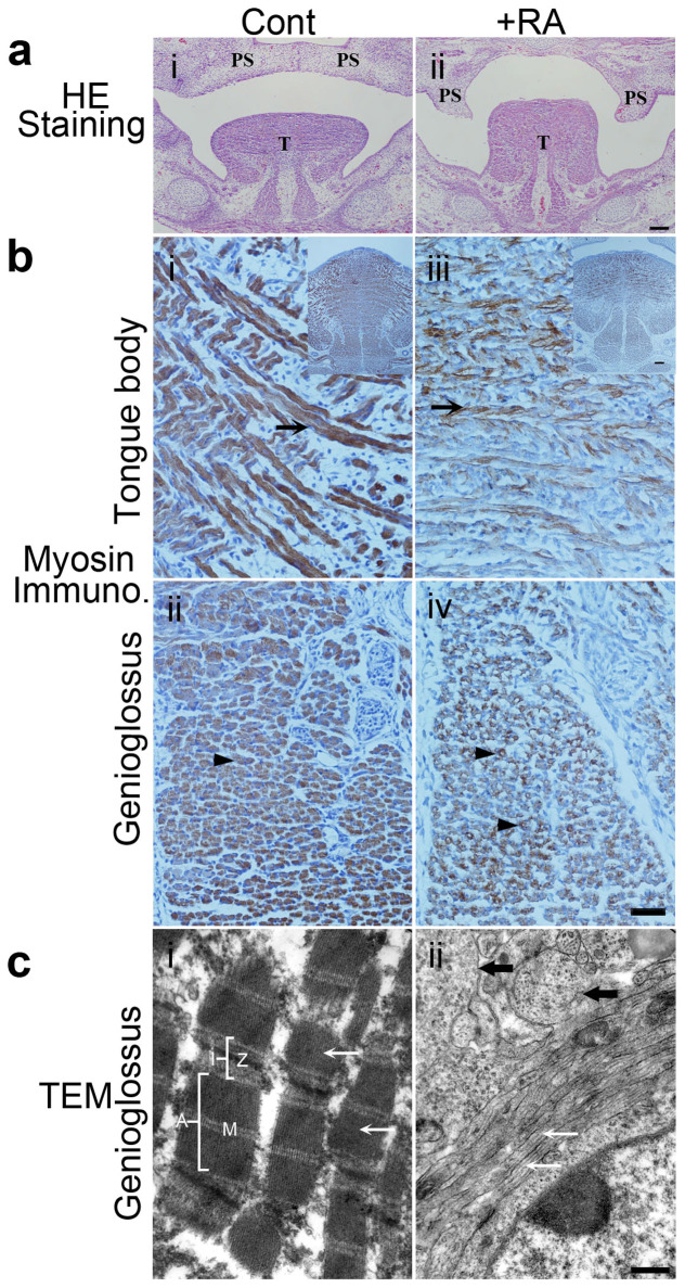
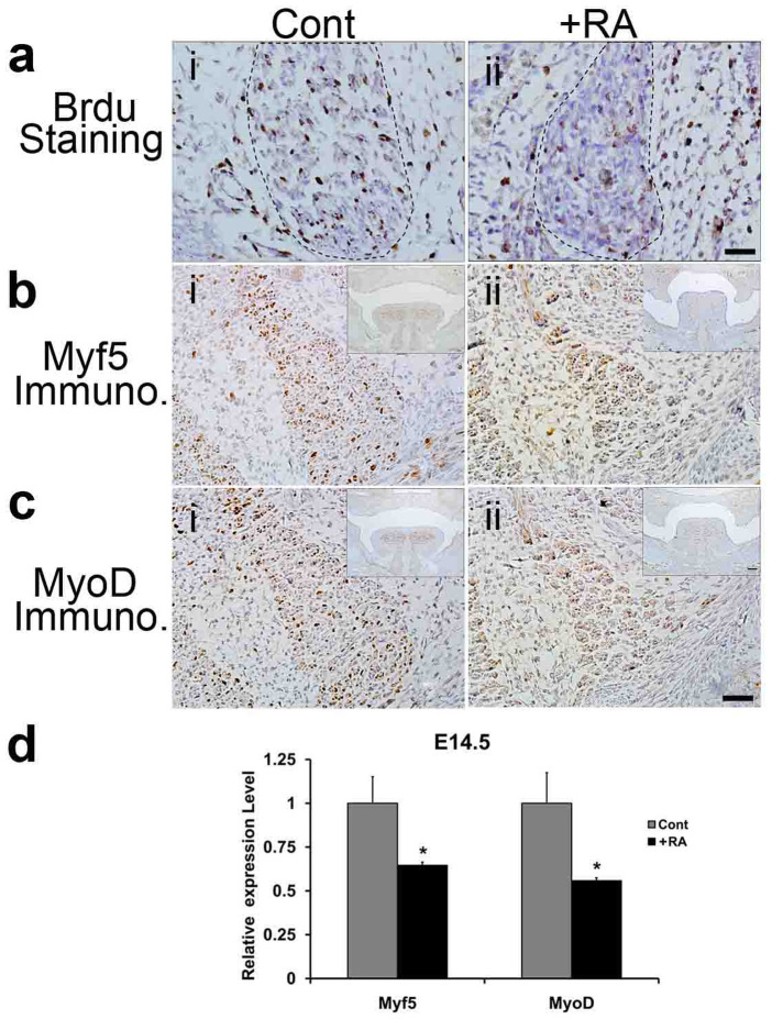
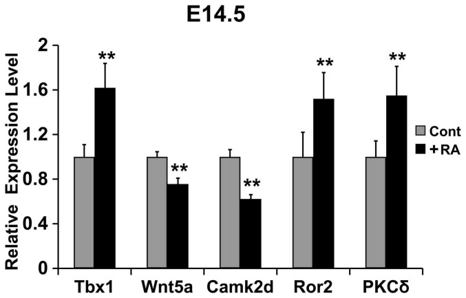
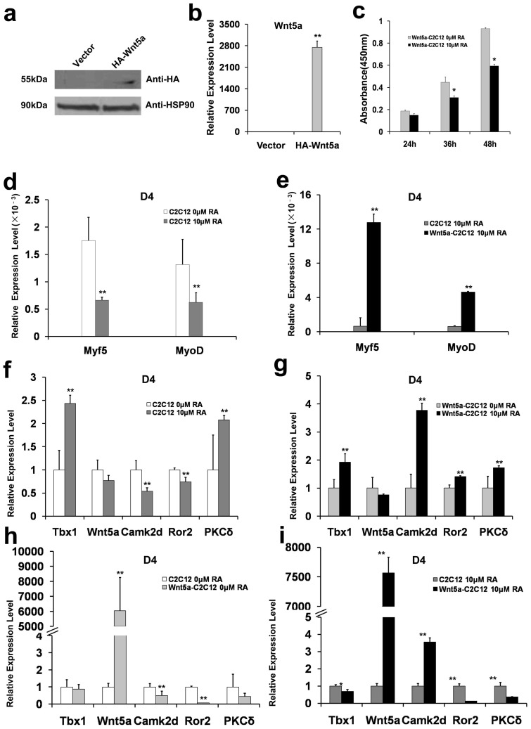
Although proper tongue development is relevant to other structures in the craniofacial region, the molecular details of muscle development in tongue remain poorly understood. Here, we report that pregnant mice treated with retinoic acid (+RA) produce embryos with tongue malformation and a cleft palate. Histological analyses revealed that at E14.5, the tongues of +RA fetuses failed to descend and flatten. Ultrastructural analysis showed that at perinatal stage E18.5, the myofilaments failed to form normal structures of sarcomeres, and arranged disorderly in the genioglossus. The proliferation and levels of myogenic determination markers (Myf5 and MyoD) and myosin in the genioglossus were profoundly reduced. Wnt5a and Camk2d expressions were down-regulated, while levels of Tbx1, Ror2, and PKCδ were up-regulated in the tongues of +RA fetuses. In mock- and Wnt5a-transfected C2C12 (Wnt5a-C2C12) cells, Wnt5a overexpression impaired proliferation, and maintained Myf5 at a relative high level after RA treatment. Furthermore, Wnt5a overexpression positively correlated with levels of Camk2d and Ror2 in C2C12 cells after RA exposure. These data support the hypothesis that the Wnt5a/CaMKII pathway is directly involved in RA-induced hypoplasia and disorder of tongue muscles.


Figures

Figure 1
Figure 1. RA-induced tongue malformation at E14.5 and E15.5, and morphology of the genioglossus at E18.5.
(a): HE staining of coronal sections of the tongue from E14.5 mouse fetus. The tongue (T) was at higher positions and bilateral palatal shelves (PS) were vertically positioned that formed a cleft in the +RA fetus (ii) versus control fetus (i). The bilateral palatal shelves (PS) in control fetus (i) already elevated above the tongue to the horizontal position and merged each other. Bar = 100 μm. (b): Myosin immunocytochemical staining of tongue muscles. Myosin expression in the tongue body (iii) and the genioglossus muscle (iv) were decreased in the +RA fetus versus control fetus. A higher magnification of the tongue intrinsic muscle from control fetuses showed multinucleated myotubes expressing high levels of myosin ((i), arrow). In +RA fetuses, myosin expression in myotubes ((iii) arrow) became weaker. In a transverse section of the genioglossus muscle, myosin was expressed at high levels in the control myotubes ((ii), arrowhead); in +RA fetuses, myosin staining was very weak. Bar = 40 μm. Upper right inserts in (i and iii) show images at lower magnifications, Bar = 100 μm. (c): Transmission electron microscopy examination of sagittal sections of the genioglossus. In control (i), myofibrils (white arrow) and sarcomeres were arranged longitudinally with integral I and A bands, and clear Z- and M-lines. In +RA (ii), the classic structures of sarcomeres of myofibrils were not found, myofilament bundles were arranged longitudinally (white arrow) and transversely (black arrow) in a single cell. Bar = 500 nm. Cont.: control mouse fetus; +RA: RA-exposed mouse fetus.PS: palatal shelves; T: tongue.
Figure 2
Figure 2. BrdU cell proliferation assay and immunohistochemistry (IHC) assay of the genioglossus at E14.5, and qRT-PCR analyses of Myf5 and MyoD expression at E14.5.
(a): Cell proliferation assay. At E14.5, the numbers of BrdU positive cells/cm2 in the genioglossus of +RA mouse fetuses (ii), decreased to 35.3% of those observed in controls (i) (n = 3 mice per group, P < 0.01). The boundary of genioglossus was delineated by the dotted line. (b)–(c): IHC assay. (b) Myf5 protein level was apparently lower in the genioglossus of the +RA mouse fetus (ii) than control (i). Bar = 40 μm. (c) MyoD protein level in the genioglossus of the +RA fetus (ii) was lower than control (i). Bar = 40 μm. Upper right inserts in (b ii and c iii) show images at lower magnifications, Bar = 100 μm. (d) qRT-PCR analyses. The mRNA levels of Myf5 and MyoD decreased significantly in the +RA fetus as compared to the controls (n = 5, P < 0.05). Cont: control mouse fetus; +RA: RA-exposed mouse fetus.
Figure 3
Figure 3. qRT-PCR analyses of the mRNA levels of Tbx1, Wnt5a, Camk2d, Ror2 and PKCδ in the tongues of E14.5 mouse fetuses.
The mRNA levels of Tbx1, Ror2 and PKCδ in the +RA fetuses increased 62%, 52% and 55%, respectively, as compared to controls; the mRNA levels of Wnt5a and Camk2d in the +RA fetuses decreased 24% and 38%, respectively, compared to controls. Experiments were performed in triplicate, and ** denotes P < 0.01.
Figure 4
Figure 4. Western blot and qRT-PCR analyses of Wnt5a expression, and qRT-PCR analyses of Myf5, MyoD and related genes expression in differentiated C2C12 cells.
(a). Western blot analysis of HA-Wnt5a expression. C2C12 cells were transfected with empty vector (Vector) or expression vector encoding the HA-tagged Wnt5a (HA-Wnt5a). HSP90 was used as a sample loading control. (b) qRT-PCR analysis of the mRNA level of HA-tagged Wnt5a. (c) Proliferation of HA-Wnt5a-C2C12 Cells with and without RA treatment. Following the indicated RA treatments, optical density at 450 nm of the HA-Wnt5a-C2C12 cells decreased to 80%, 69% and 63% of the control cells, at the time intervals of 24 h, 36 h, and 48 h, respectively. The changes at 36 h and 48 h were statistically significant (n = 3, *P < 0.05). (d) Differentiated C2C12 cells were treated with or without RA (10 μM), and the mRNA levels of Myf5 and MyoD were determined by qRT-PCR at day 4 (D4). (e) Differentiated control C2C12 cells and HA-Wnt5a-C2C12 cells were treated with 10 μM RA, and the mRNA levels of Myf5 and MyoD were determined using qRT-PCR (D4). (f) Differentiated C2C12 cells were incubated with or without RA (10 μM), and the mRNA levels of Tbx1, Wnt5a, Camk2d, Ror2 and PKCδ were determined by qRT-PCR (D4). (g) Differentiated HA-Wnt5a C2C12 cells were treated with or without RA (10 μM), and the mRNA levels of Tbx1, Wnt5a, Camk2d, Ror2 and PKCδ were measured using qRT-PCR (D4). (h)–(i): qRT-PCR analyses of expression levels of Tbx1, Wnt5a, Cam2d, Ror2, and PKCδ in control C2C12 cells and HA-Wnt5a C2C12 cells (D4) under basal condition (h) and after RA (10 μM) treatment (i). All experiments were performed in triplicate, and ** denotes P < 0.01. In (a), the full length blots were presented in Supplementary Figure S3.

References

    1. Hall J. G. Importance of muscle movement for normal craniofacial development. J Craniofac Surg. 21, 1336–1338 (2010). - PubMed
    1. Yamane A. et al. Expression of myogenic regulatory factors during the development of mouse tongue striated muscle. Arch Oral Biol. 45, 71–78 (2000). - PubMed
    1. Dalrymple K. R., Prigozy T. I., Mayo M., Kedes L. & Shuler C. F. Murine tongue muscle displays a distinct developmental profile of MRF and contractile gene expression. Int J Dev Biol. 43, 27–37 (1999). - PubMed
    1. Mok G. F. & Sweetman D. Many routes to the same destination: lessons from skeletal muscle development. Reproduction. 141, 301–312 (2011). - PubMed
    1. Duester G. Retinoic acid synthesis and signaling d uring early organogenesis. Cell. 134, 921–931 (2008). - PMC - PubMed

Publication types

MeSH terms