. 2014 Oct 7;130(15):1262-73.
doi: 10.1161/CIRCULATIONAHA.114.006185. Epub 2014 Aug 14.

Cardiac CaM Kinase II genes δ and γ contribute to adverse remodeling but redundantly inhibit calcineurin-induced myocardial hypertrophy

Affiliations

Cardiac CaM Kinase II genes δ and γ contribute to adverse remodeling but redundantly inhibit calcineurin-induced myocardial hypertrophy

Michael M Kreusser et al. Circulation. .

Abstract

Background: Ca(2+)-dependent signaling through CaM Kinase II (CaMKII) and calcineurin was suggested to contribute to adverse cardiac remodeling. However, the relative importance of CaMKII versus calcineurin for adverse cardiac remodeling remained unclear.

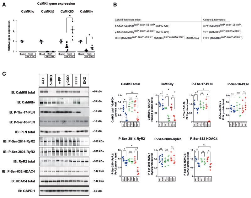
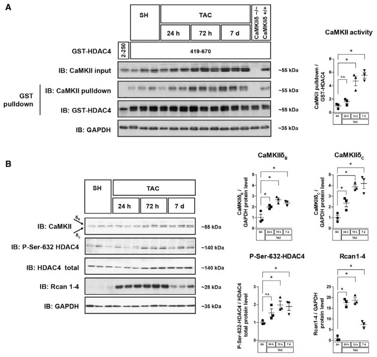
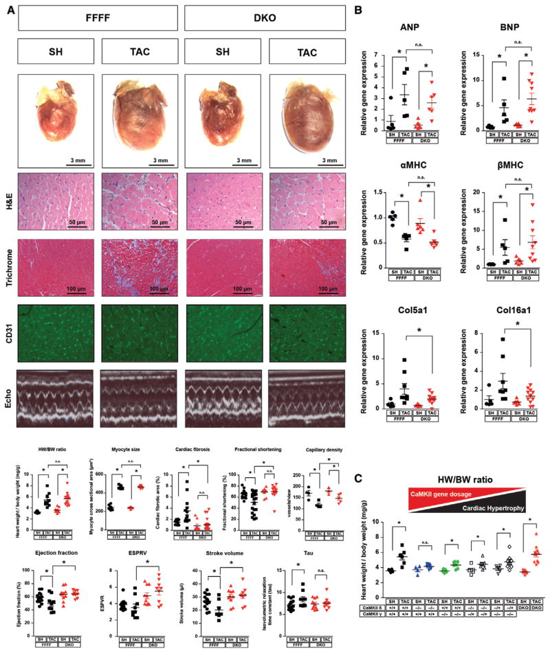
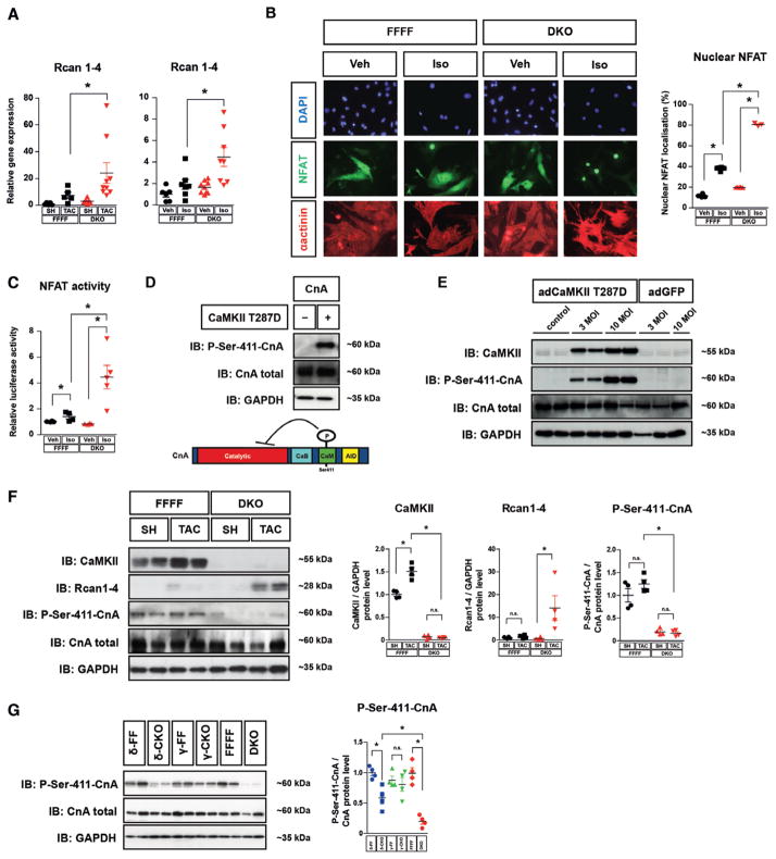
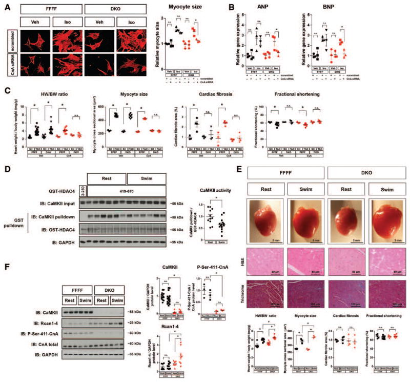
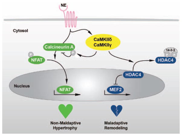
Methods and results: We generated double-knockout mice (DKO) lacking the 2 cardiac CaMKII genes δ and γ specifically in cardiomyocytes. We show that both CaMKII isoforms contribute redundantly to phosphorylation not only of phospholamban, ryanodine receptor 2, and histone deacetylase 4, but also calcineurin. Under baseline conditions, DKO mice are viable and display neither abnormal Ca(2+) handling nor functional and structural changes. On pathological pressure overload and β-adrenergic stimulation, DKO mice are protected against cardiac dysfunction and interstitial fibrosis. But surprisingly and paradoxically, DKO mice develop cardiac hypertrophy driven by excessive activation of endogenous calcineurin, which is associated with a lack of phosphorylation at the auto-inhibitory calcineurin A site Ser411. Likewise, calcineurin inhibition prevents cardiac hypertrophy in DKO. On exercise performance, DKO mice show an exaggeration of cardiac hypertrophy with increased expression of the calcineurin target gene RCAN1-4 but no signs of adverse cardiac remodeling.

Conclusions: We established a mouse model in which CaMKII's activity is specifically and completely abolished. By the use of this model we show that CaMKII induces maladaptive cardiac remodeling while it inhibits calcineurin-dependent hypertrophy. These data suggest inhibition of CaMKII but not calcineurin as a promising approach to attenuate the progression of heart failure.

Keywords: CaMKII; calcineurin; cardiac hypertrophy; heart failure; signal transduction.


Figures

Figure 1
Figure 1
CaMKIIδ and CaMKIIγ contribute redundantly to cardiac CaMKII activity. A, Real-time polymerase chain reaction analysis of cardiac CaMKII isoforms 3 weeks after sham (SH) or transverse aortic constriction (TAC) surgery in wild-type mice. The relative expression levels were normalized to CaMKII in brain tissue. All values are reported as mean±SEM (n≥3, *P<0.05). B, To generate cardiomyocyte-specific conditional CaMKIIδ and γ single knockout (δ-CKO and γ-CKO) and CaMKIIδ/CaMKIIγ double knockout mice (DKO), CaMKIIδloxP-exon1/2-loxP (δ-FF), CaMKIIγloxP-exon1/2-loxP (γ-FF), and CaMKIIδloxP-exon1/2-loxP, CaMKIIγloxP-exon1/2-loxP (FFFF) mice were crossed to transgenic mice harboring Cre-recombinase under the control of the αMHC promoter. C, Western blot analysis using antibodies directed against CaMKII, CaMKIIγ, total PLB, phospho-PLB-Thr17, phospho-PLB-Ser16, total RyR2, phospho-RyR2-Ser2814, phospho-RyR2-Ser2808, total HDAC4, phospho-HDAC4-Ser632, and GAPDH (as loading control). Left ventricular extracts from δ-CKO, γ-CKO and DKO mice and their Cre-negative littermates (δ-FF, γ-FF, and FFFF) were analyzed (n=4 per group). Quantitative analysis of the expression and phosphorylation of the proteins is shown at right. All values are presented as mean±SEM. *P<0.05. n.s. indicates not significant.
Figure 2
Figure 2
Calcineurin precedes CaMKII activation on pathological pressure overload. Western blot analyses were performed in cardiac extracts from wild-type mice. Organs were harvested at different time points after transverse aortic constriction (TAC) surgery as indicated. A, Western blot analysis after GST-HDAC4 pulldown using GST-HDAC4 419 to 670, which contains a CaMKII-activity dependent binding site. GST-HDAC4 2 to 250, which lacks a CaMKII binding domain, served as a negative control. As an additional negative control, protein lysates from global CaMKIIδ mice vs wild-type mice were used. GST-HDAC4 input was visualized by GST-immunoblotting, associated endogenous CaMKII is visualized by CaMKII-immunoblotting. The degree of CaMKII binding to GST-HDAC4 419-670 is a specific measure for CaMKII activity. A quantification of CaMKII binding normalized to the input of GST-HDAC4 is shown. B, Western blot analysis using antibodies directed against CaMKII, HDAC4, phospho-HDAC4-Ser632, RCAN1-4, and GAPDH. Quantitative analysis of the expression and phosphorylation of the proteins is shown. For all experiments n≥3 per group were used. All values are presented as mean±SEM. *P<0.05. n.s. indicates not significant.
Figure 3
Figure 3
Dissociation of cardiac dysfunction and interstitial fibrosis from myocardial hypertrophy. A, Double knockout (DKO) and CaMKIIδloxP-exon1/2-loxP, CaMKIIγloxP-exon1/2-loxP (FFFF) mice were randomized to either transverse aortic constriction (TAC) or sham (SH) surgery and euthanized after 3 weeks. Representative images of the total hearts, H&E, Masson trichrome, and CD31 staining, echocardiographic M-modes, and quantification of heart weight/body weight ratios, myocyte size, fibrosis area, capillary density, and fractional shortening are shown (n≥8 per group). Values of left ventricular ejection fraction, isovolumetric relaxation time constant (Tau), end systolic pressure volume relation (ESPVR), and stroke volume measured in a working heart preparation are shown (n≥7 per group). B, Fold-changes in mRNA levels of the hypertrophic markers ANP, BNP, αMHC, and βMHC and fold-changes in collagen expression (n≥5 per group). C, Heart weight/body weight ratios of SH- and TAC-operated mice with the indicated genotypes (n≥4 per group). All values are presented as mean±SEM. *P<0.05. n.s. indicates not significant.
Figure 4
Figure 4
CaMKII controls calcineurin A (CnA) activity. A, Real-time-PCR analysis of RCAN1-4 in hearts from animals with genotypes and treatments as indicated (≥5 per group). B and C, Neonatal mouse ventricular myocytes (NMVMs) from double knockout (DKO) and CaMKIIδloxP-exon1/2-loxP, CaMKIIγloxP-exon1/2-loxP (FFFF) mice were used for cell-based experiments. B, NFAT-GFP was adenovirally expressed in NMVMs. Cells were stained for α-actinin (shown in red) and with DAPI nuclear stain (shown in blue). The percentage of cells in which NFAT-GFP was localized to the nucleus is indicated (>100 cardiomyocytes per well; n≥3 per condition). C, NFAT-luciferase reporter assay in NMVMs (n≥3 per condition). D, Western blot analysis was performed using lysates from COS cells that were transfected with CnA in the absence and presence of constitutive active CaMKIIδ (CaMKII-T287D). A schematic diagram of CnA indicates the position of the CaMKII phosphorylation site at Ser411, which is located within the calmodulin-binding domain of CnA (AID indicates autoinhibitory domain; CaM, calmodulin-binding domain; CnB, calcineurin B-binding domain). E, Adenoviral overexpression of CaMKII-T287D (adCaMKII-T287D) in neonatal rat ventricular myocytes (NRVMs) results in hyper-phosphorylation of CnA-Ser411 compared with noninfected (control) NRVMs and NRVMs infected with GFP. F, Western blot analysis of total CaMKII, RCAN1-4, total CnA, and CnA-Ser411 in cardiac extracts from FFFF and DKO mice 3 weeks after transverse aortic constriction (TAC) or sham (SH) surgery (n≥3 per group). G, Western blot analysis of total CnA and CnA-Ser411 in δ-CKO, γ-CKO or DKO vs their Cre-negative littermates (n=4 per group). For all Western blot experiments, GAPDH was used as loading control. Quantitative analysis of the expression and phosphorylation of the proteins is shown at right (F and G). All values are presented as mean±SEM. *P<0.05. n.s. indicates not significant.
Figure 5
Figure 5
Cardiac hypertrophy but not dysfunction is controlled by calcineurin during pathological and physiological hypertrophy in double knockout mice (DKO). A and B, Neonatal mouse ventricular myocytes (NMVMs) from CaMKIIδloxP-exon1/2-loxP, CaMKIIγloxP-exon1/2-loxP (FFFF) and DKO mice were transfected with two siRNAs directed against CnA or with scrambled siRNA (knockdown efficiency is shown in Figure VA in the online-only Data Supplement). NMVMs were then treated with Iso or vehicle. A, ANP and BNP gene expression in NMVMs after Iso-stimulation (n≥3 per condition). B, Cells were stained for α-actinin (shown in red) and cardiomyocyte size was determined after Iso-stimulation (>100 cardiomyocytes per well; n≥3 per condition). C, FFFF and DKO mice underwent SH or TAC surgery and were treated with cyclosporine A (CyA; 4 mg/Kg/d, 3 weeks IP; n≥7) or vehicle (n≥3) as indicated. CyA treatment was started at the day of transverse aortic constriction (TAC) or sham (SH) surgery. Three weeks later the mice were euthanized. Heart weight/body weight ratios, myocyte cross sectional areas, quantification of cardiac fibrosis from Masson trichrome–stained myocardial sections, and fractional shortening assessed by echocardiography are shown. D–F, FFFF and DKO mice were randomly assigned to a 14-day swimming or resting group, respectively. D, Cardiac CaMKII activity was determined in heart lysates by GST-HDAC4 pulldown using GST-HDAC4 419-670 (GST-HDAC4 2–250 served as specificity control). A quantification of CaMKII-activity dependent binding as measure for CaMKII activity is given (n≥11 per group). E, Representative images of whole hearts as well as H&E and Masson trichrome staining of left ventricular wall sections. Quantification of heart weight/body weight ratios, myocyte size, fibrosis area, and fractional shortening (n≥3 per group). F, Western blot analysis using antibodies directed against CaMKII, total CnA, phospho-CnA-Ser411, RCAN1-4, and GAPDH. Quantitative analysis of the expression and phosphorylation of the proteins is shown (n≥7 per group). All values are presented as mean±SEM. *P<0.05. n.s. indicates not significant.
Figure 6
Figure 6
Schematic model. Stimulation of β-adrenergic receptors by norepinephrine (NE) leads to an activation of the 2 Ca2+-dependent pathways calcineurin and CaMKII (mediated by the isoforms CaMKIIδ and CaMKIIγ). Activated CaMKII phosphorylates HDAC4, which leads to a translocation of HDAC4 from the nucleus to the cytosol. Thereby, the repressive effect of HDAC4 on transcription factors as myocyte enhancer factor 2 (MEF2) is interrupted and a maladaptive remodeling gene program is initiated. As a second mechanism, CaMKII phosphorylates calcineurin A (CnA), which leads to repression of the calcineurin-NFAT pathway and thereby acts antihypertrophic. Unexpectedly, the endogenous calcineurin-NFAT driven prohypertrophic pathway is not maladaptive.

References

    1. Vitali E, Colombo T, Bruschi G, Garatti A, Russo C, Lanfranconi M, Frigerio M. Different clinical scenarios for circulatory mechanical support in acute and chronic heart failure. Am J Cardiol. 2005;96(12A):34L–41L. - PubMed
    1. Backs J, Olson EN. Control of cardiac growth by histone acetylation/deacetylation. Circ Res. 2006;98:15–24. - PubMed
    1. Zarain-Herzberg A, Fragoso-Medina J, Estrada-Avilés R. Calcium-regulated transcriptional pathways in the normal and pathologic heart. IUBMB Life. 2011;63:847–855. - PubMed
    1. Colomer JM, Mao L, Rockman HA, Means AR. Pressure overload selectively up-regulates Ca2+/calmodulin-dependent protein kinase II in vivo. Mol Endocrinol. 2003;17:183–192. - PubMed
    1. Hoch B, Meyer R, Hetzer R, Krause EG, Karczewski P. Identification and expression of delta-isoforms of the multifunctional Ca2+/calmodulin-dependent protein kinase in failing and nonfailing human myocardium. Circ Res. 1999;84:713–721. - PubMed

Publication types

MeSH terms

Substances