Review
. 2014 Sep-Oct;3(5):301-30.
doi: 10.1002/wdev.138. Epub 2014 May 29.

Maternal control of the Drosophila dorsal-ventral body axis

Affiliations
Review

Maternal control of the Drosophila dorsal-ventral body axis

David S Stein et al. Wiley Interdiscip Rev Dev Biol. 2014 Sep-Oct.

Abstract

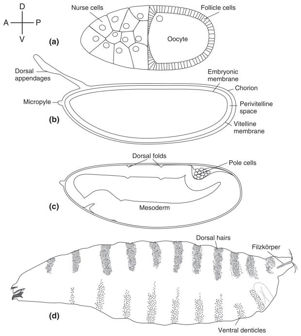
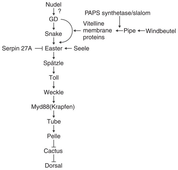
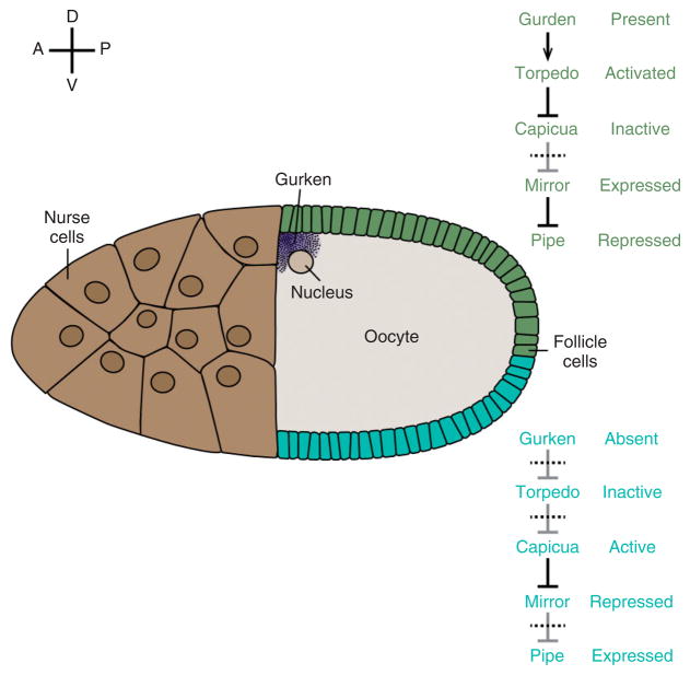
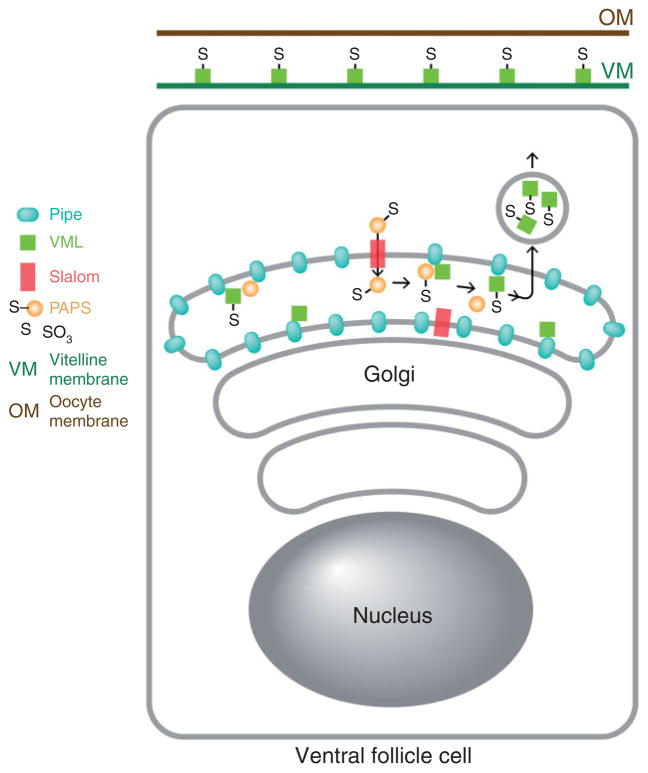
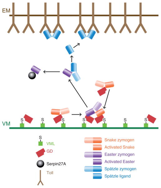
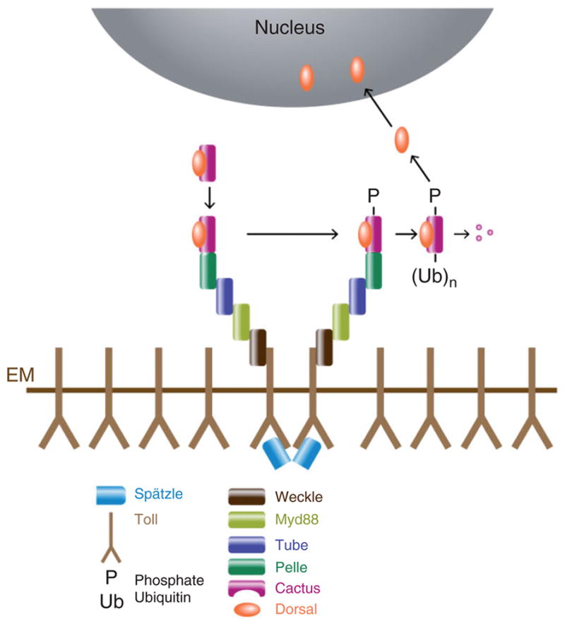
The pathway that generates the dorsal-ventral (DV) axis of the Drosophila embryo has been the subject of intense investigation over the previous three decades. The initial asymmetric signal originates during oogenesis by the movement of the oocyte nucleus to an anterior corner of the oocyte, which establishes DV polarity within the follicle through signaling between Gurken, the Drosophila Transforming Growth Factor (TGF)-α homologue secreted from the oocyte, and the Drosophila Epidermal Growth Factor Receptor (EGFR) that is expressed by the follicular epithelium cells that envelop the oocyte. Follicle cells that are not exposed to Gurken follow a ventral fate and express Pipe, a sulfotransferase that enzymatically modifies components of the inner vitelline membrane layer of the eggshell, thereby transferring DV spatial information from the follicle to the egg. These ventrally sulfated eggshell proteins comprise a localized cue that directs the ventrally restricted formation of the active Spätzle ligand within the perivitelline space between the eggshell and the embryonic membrane. Spätzle activates Toll, a transmembrane receptor in the embryonic membrane. Transmission of the Toll signal into the embryo leads to the formation of a ventral-to-dorsal gradient of the transcription factor Dorsal within the nuclei of the syncytial blastoderm stage embryo. Dorsal controls the spatially specific expression of a large constellation of zygotic target genes, the Dorsal gene regulatory network, along the embryonic DV circumference. This article reviews classic studies and integrates them with the details of more recent work that has advanced our understanding of the complex pathway that establishes Drosophila embryo DV polarity. For further resources related to this article, please visit the WIREs website.

Conflict of interest: The authors have declared no conflicts of interest for this article.


Conflict of interest statement

Conflict of interest: The authors have declared no conflicts of interest for this article.

Figures

FIGURE 1
FIGURE 1
Drosophila dorsal–ventral polarity from the oocyte to the first instar larva. The compass at the upper left indicates the direction of Anterior (A), Posterior (P), Dorsal (D), and Ventral (V) for each schematic drawing. Relevant structures are labeled. (a) Stage 10 oocyte. (b) Egg contained within the eggshell. (c) Embryo undergoing germband extension during gastrulation. (d) Cuticle of first instar larva.
FIGURE 2
FIGURE 2
Wild-type and dorsalized embryonic phenotypes. (a) Cuticle of an embryo from a wild-type female. Anterior is to the left and ventral is on the bottom. (b) Dorsalized cuticle of a larva from a gdVM90/gdVM90 mutant female.
FIGURE 3
FIGURE 3
The current understanding of the order of action and epistatic relationships of the gene products known to be involved in dorsal–ventral patterning of the embryo.
FIGURE 4
FIGURE 4
Model for the ventrally restricted expression of pipe in the follicle cell layer. Schematic drawing of a stage 10 oocyte. pipe is expressed in the ventral region (blue) but repressed in the dorsal epithelium (green). Relevant effector molecules and their activation states in ventral and dorsal follicle cells are indicated at right.
FIGURE 5
FIGURE 5
Sequential asymmetry along the DV axis of polarity from the oocyte to zygotic gene expression in the embryo. In all panels, anterior is to the left and ventral is down. (a, b) Stage 10 oocyte showing the dorsal anterior localization of gurken mRNA (a) and ventrally restricted expression of pipe mRNA (b). (c–e) Syncytial blastoderm embryos showing ventral localization of GD-GFP after injection into the pervitelline space (c), ventral-to-dorsal gradient of Cactus-LacZ degradation visualized by X-gal staining (d), and ventral-to-dorsal gradient of Dorsal nuclear localization visualized with anti-Dorsal antibody (e). (f) Cellular blastoderm embryo showing the expression domain of the mRNA encoding the Dorsal target gene twist in the presumptive mesoderm.
FIGURE 6
FIGURE 6
Diagram depicting the sulfation of VML by Pipe in a ventral follicle cell. PAPS is transported from its cytoplasmic site of synthesis into the Golgi apparatus by Slalom. Pipe present in the Golgi lumen transfers sulfate from PAPS to VML. Sulfated VML is then secreted and incorporated into the vitelline membrane layer of the eggshell.
FIGURE 7
FIGURE 7
Diagram depicting the formation of the Spätzle ligand in the ventral perivitelline space. VML-associated and free GD processes and activates Snake. Only GD that is associated with Pipe-sulfated VML promotes the interaction between activated Snake and the Easter zymogen, which leads to Easter cleavage and activation. Activated Easter then processes and converts Spätzle into the active Toll ligand. Active Easter is bound by and inactivated by Serpin 27A.
FIGURE 8
FIGURE 8
Diagram depicting the components that mediate Toll activation and signaling, leading to Dorsal nuclear uptake. Binding of activated Spätzle to Toll leads to the recruitment of a complex comprised of Myd88, Tube, and Pelle, in a process that depends upon Weckle. This leads to the phosphorylation, ubiquitination, and degradation of Cactus, releasing Dorsal to enter the nucleus.
FIGURE 9
FIGURE 9
Different levels of nuclear Dorsal result in differential gene expression. (a) Diagram depicting the nuclear Dorsal gradient. Colors on the outside circle indicate the regions in which zygotic genes classified as Type I, II, or III targets exhibit Dorsal-dependent transcriptional regulation. Type I targets are transcribed in the ventral mesodermal region. Type II targets are transcribed in the lateral neuroectoderm region and repressed in the mesodermal anlagen. Type III targets transition from either a repressed (ventral/lateral) to a de-repressed (dorsal) state, or from an active (lateral) to an inactive (dorsal) state, with the transition point occurring within the dorsal half of the embryo. (b) Cross-section of an embryo used for in situ hybridization to visualize the expression domains of snail (sna) (Type I), ventral nervous system defective (vnd) (Type II) and intermediate neuroblasts defective (ind) (Type II), short gastrulation (sog) (Type III) and decapentaplegic (dpp) (Type III). (Reprinted with permission from Ref . Copyright 2009 Cold Spring Harbor Press)
FIGURE 10
FIGURE 10
A model for Dorsal-mediated control of the snail (sna) transcription unit in the mesoderm and in the dorsal ectoderm/neurectoderm. At top is shown the position of the sna transcription unit in relation to the positions of the primary and shadow enhancer regions. In regions of lower nuclear Dorsal concentrations (dorsal ectoderm/neurectoderm), there is no Dorsal binding to either enhancer, and RNA Pol II is paused at the start site., In the ventral cells with high levels of nuclear Dorsal (mesoderm), Dorsal is bound to both enhancers. The binding of Dorsal induces the previously paused RNA Pol II to initiate sna transcription.

References

    1. González-Reyes A, St Johnston D. Role of oocyte position in establishment of anterior-polarity polarity in Drosophila. Science. 1994;266:639–642. doi: 10.1126/science.7939717. - DOI - PubMed
    1. Montell DJ, Keshishian H, Spradling AC. Laser ablation studies of the role of the Drosophila oocyte nucleus in pattern formation. Science. 1991;254:290–293. doi: 10.1126/science.1925585. - DOI - PubMed
    1. Roth S, Neuman-Silberberg FS, Barcelo G, Schüpbach T. cornichon and the EGF receptor signaling process are necessary for both anterior-posterior and dorsal-ventral pattern formation in Drosophila. Cell. 1995;81:967–978. doi: 10.1016/0092-8674(95)90016-0. - DOI - PubMed
    1. Roth S, Jordan P, Karess R. Binuclear Drosophila oocytes: consequences and implications for dorsal-ventral patterning in oogenesis and embryogenesis. Development. 1999;126:927–934. - PubMed
    1. Nüsslein-Volhard C. Maternal effect mutations that alter the spatial coordinates of the embryo of Drosophila melanogaster. In: Subtelny S, Konigsberg IR, editors. Determinants of Spatial Organization: the Thirty-Seventh Symposium of the Society for Developmental Biology. New York: Academic Press; 1979. pp. 185–211.

FURTHER READING

    1. Flybase; St Pierre SE, Ponting L, Stefancsik R, McQuilton P the FlyBase Consortium. Flybase 102 - advanced approaches to interrogating Flybase. Nucleic Acids Res. 2014;42(D1):D780–D788. doi: 10.1093/nar/gkt1092. Available at: http://www.flybase.org. - DOI - PMC - PubMed
    1. The interactive fly. [Accessed May 19, 2014];Brody TB. 2013 Available at: http://www.sdbonline.org/sites/fly/aimain/1aahome.htm.
    1. Gilmore TD, Wolenski FS. NF-κB: where did it come from and why? Immunol Rev. 2012;246:14–35. doi: 10.1111/j.1600-065X.2012.01096.x. - DOI - PubMed
    1. Lynch JA, Roth S. The evolution of dorsal-ventral patterning mechanisms in insects. Genes Dev. 2011;25:107–118. doi: 10.1101/gad.2010711. - DOI - PMC - PubMed
    1. Valanne S, Wang JH, Rämet M. The Drosophila Toll signaling pathway. J Immunol. 2011;186:649–656. doi: 10.4049/jimmunol.1002302. - DOI - PubMed

Publication types

LinkOut - more resources