. 2014 Sep;24(9):1091-107.
doi: 10.1038/cr.2014.108. Epub 2014 Aug 15.

Hydrogen peroxide primes heart regeneration with a derepression mechanism

Affiliations

Hydrogen peroxide primes heart regeneration with a derepression mechanism

Peidong Han et al. Cell Res. 2014 Sep.

Abstract

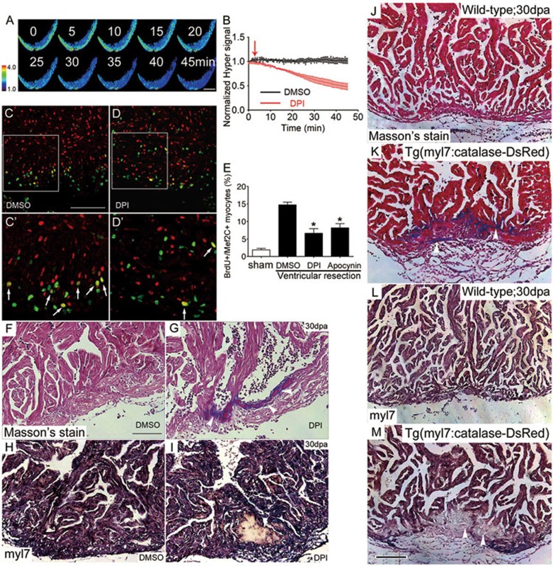
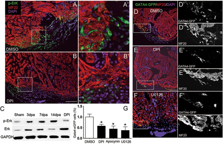
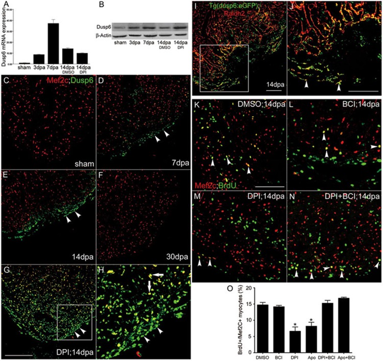
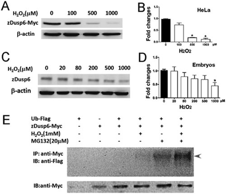
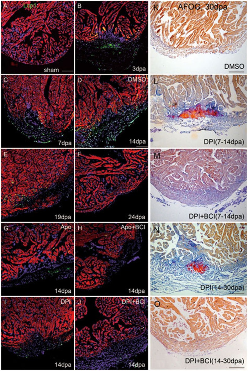
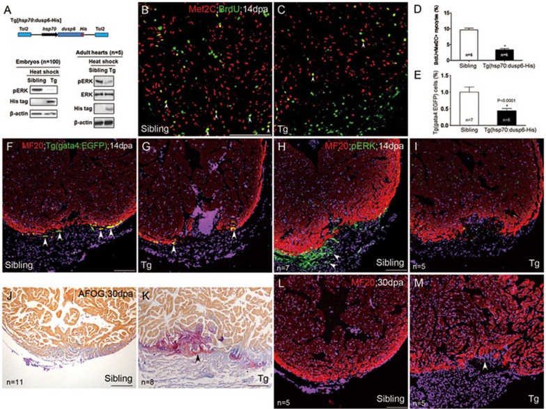
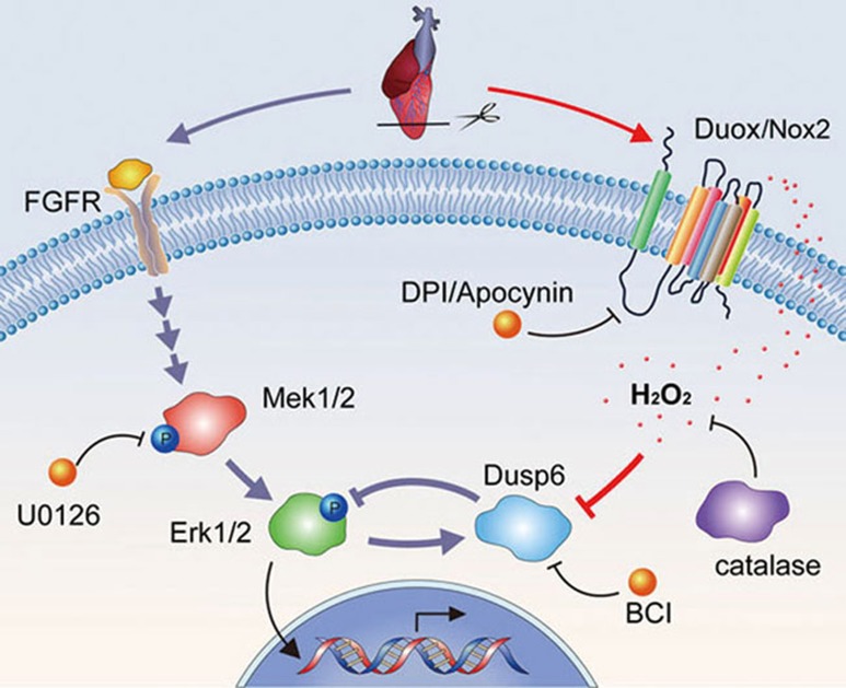
While the adult human heart has very limited regenerative potential, the adult zebrafish heart can fully regenerate after 20% ventricular resection. Although previous reports suggest that developmental signaling pathways such as FGF and PDGF are reused in adult heart regeneration, the underlying intracellular mechanisms remain largely unknown. Here we show that H2O2 acts as a novel epicardial and myocardial signal to prime the heart for regeneration in adult zebrafish. Live imaging of intact hearts revealed highly localized H2O2 (~30 μM) production in the epicardium and adjacent compact myocardium at the resection site. Decreasing H2O2 formation with the Duox inhibitors diphenyleneiodonium (DPI) or apocynin, or scavenging H2O2 by catalase overexpression markedly impaired cardiac regeneration while exogenous H2O2 rescued the inhibitory effects of DPI on cardiac regeneration, indicating that H2O2 is an essential and sufficient signal in this process. Mechanistically, elevated H2O2 destabilized the redox-sensitive phosphatase Dusp6 and hence increased the phosphorylation of Erk1/2. The Dusp6 inhibitor BCI achieved similar pro-regenerative effects while transgenic overexpression of dusp6 impaired cardiac regeneration. H2O2 plays a dual role in recruiting immune cells and promoting heart regeneration through two relatively independent pathways. We conclude that H2O2 potentially generated from Duox/Nox2 promotes heart regeneration in zebrafish by unleashing MAP kinase signaling through a derepression mechanism involving Dusp6.


Figures

Figure 1
Figure 1
Epicardial Duox-generated H2O2 associated with myocardial regeneration. (A-C) After imaging of Tg(tcf21:nucEGFP), we performed RNA in situ hybridization with duox probes on 10-μm cryosections of Tg(tcf21:nucEGFP) hearts at sham, 7 and 14 dpa. Note the induced expression of duox and its colocalization with epicardial reporter Tg(tcf21:nucEGFP) in injured areas (arrowheads) at 7 and 14 dpa (B, C) while little staining at sham (A). Scale bar for A-C, 50 μm. (D) Cardiac-specific expression of Hyper in a zebrafish embryo. Hyper was restricted in the Tg(myl7:Hyper) transgenic heart at 48 hpf. (E-H) Ex vivo Hyper heart images from sham control (E), 7 dpa (F), 14 dpa (G) and 30 dpa (H). Spatially resolved H2O2 image, indexed by the ratio between the F488 and F405 images of Hyper (left panels), is presented in pseudocolor. (I) Ratiometric Hyper signals (F488/F405) averaged over the regenerative zone of injured heart during the first month of regeneration after resection. n = 3. (J) Representative transmural spatial profiles of the Hyper signal at 14 dpa (red line) and sham (green line) hearts. (K) Ex vivo calibration of Hyper F488/F405 ratio as a function of ambient H2O2 concentration. n = 3. Arrow denotes the average F488/F405 ratio seen in regenerative zones at 14 dpa. (L-O) Redox signal showing greater epicardial and myocardial oxidization during regeneration. A 14 dpa Tg(myl7:hyper) heart was labeled with Redox sensor cc-1. Images show Hyper signals at 488 (L) and 405 nm (M), and Redox sensor cc-1 signals at 555 nm excitation (N), and the merging of L and N (O). Note that the Redox sensor cc-1 signal (arrowheads), indicative of intracellular oxidization, was most conspicuous in epicardial cells lacking Hyper expression, and also overlapped with the Hyper signal (arrows) in adjacent myocardium. Scale bars, 100 μm.
Figure 2
Figure 2
H2O2 signaling is required for heart regeneration. (A-B) Inhibiting Duox/Nox NADPH oxidases by DPI decreased H2O2 generation. Time-lapse confocal images (A) and statistics (B) of the Hyper ratio in Tg(myl7:Hyper) hearts prior to and after application of DPI (10 μM). DMSO (0.1%) was used as control. n = 3-5. (C-E) Treatment with DPI or apocynin impaired cardiac myocyte regeneration. Proliferating myocytes were identified by double-staining with anti-Mef2C (red) and anti-BrdU (green) (C', D', arrows). Note that there were fewer double-positive cells (yellow) in DPI-treated heart (D, D') than in DMSO control heart (C, C'). Quantitative results with sham, DMSO, DPI or apocynin treatment are shown in E. n = 5 to 7. See Materials and Methods for details of treatment. (F-I) Accumulated fibrin/collagens (white arrowheads in G, Masson's staining) and compromised myocardial regeneration (white arrowhead in I, in situ hybridization with myl7 probes) after DPI treatment, compared with DMSO control (F, H). (J-M) Cardiac-specific overexpression of catalase retarded heart regeneration. Tg(myl7:Catalase-DsRed) heart displayed larger amounts of fibrin/collagens (white arrowheads in K, Masson's staining) and compromised myocardial regeneration (white arrowheads in M, in situ hybridization with myl7 probes), as compared with respective non-transgenic sibling hearts at 30 dpa (J, L). n = 5-8. Scale bars, 100 μm.
Figure 3
Figure 3
H2O2 activates Erk1/2 for myocardial regeneration. (A-B) Phosphorylation of Erk1/2 (pErk1/2) in epicardial cells (A, A') in 14 dpa heart, as detected by anti-pErk1/2 antibody. Decrease of pErk1/2-positive cells by DPI treatment (B, B'). Scale bar for A and B, 100 μm. (C) Western blot showing greater pErk1/2 in injured hearts at 3, 7 and 14 dpa, and its blockade by DPI treatment at 14 dpa. (D-F) Gata4+ myocardial cells in injured areas in control Tg(gata4:EGFP) heart (DMSO treatment, D, D', D'') and their disappearance after inhibition of Duox (DPI treatment, E, E', E”), or Mek1/2 (U0126 treatment, F, F', F”). Cardiomyocytes were co-labeled with anti-EGFP and anti-MF20 antibodies and nuclei were labeled by DAPI. Scale bar for D-F, 100 μm. (G) Statistics of Gata4+ cardiomyocytes at 14 dpa.
Figure 4
Figure 4
Suppressing Dusp6 by H2O2 or BCI promotes cardiac regeneration. (A) Quantitative RT-PCR showing induced dusp6 expression during regeneration. Note that dusp6 mRNA expression was unaffected by DPI treatment. n = 5. (B) Western blotting of Dusp6 proteins at 3, 7 and 14 dpa. Note that DPI treatment further augmented Dusp6 abundance at 14 dpa. (C-F) Dusp6 immunostaining in sham control (C) and at 7, 14 and 30 dpa (D-F). (G-H) DPI treatment induced widespread intense Dusp6 protein expression in the epicardium (arrowheads) and myocardium (arrows) at 14 dpa. Similar results were obtained in apocynin-treated hearts (data not shown). (I-J) Immunostaining showing colocalization of reporter EGFP (green) in Tg(dusp6:EGFP) heart and the epicardial and endocardial marker Raldh2 protein (red) in the injured area at 14 dpa. White arrowheads in J: epicardial cells positive for both Raldh2 and EGFP. (K-N) DPI treatment diminished proliferating myocytes identified as Mef2C (red) and BrdU (green) double-positive cells at 14 dpa (M) as compared to DMSO control (K). This inhibitory effect was reversed by BCI treatment (N), while BCI alone exerted no significant effect (L). (O) Statistics of the BrdU+/Mef2C+ proliferating myocytes. Scale bars, 100 μm.
Figure 5
Figure 5
H2O2 induces ubiquitination and proteasome degradation of zebrafish Dusp6. (A-B) Exogenous H2O2 decreased the expression level of Myc-tagged zDusp6 in a dose-dependent manner in HeLa cells as evidenced by western blotting (A). The statistics of A is shown in B. β-actin was used for normalizing protein loading. (C-D) Exogenous H2O2 decreased the expression level of endogenous zDusp6 in a dose-dependent manner in zebrafish embryos at 24 hpf (C), and the statistics of C is shown in D. β-actin was used for normalizing protein loading. (E) Ub-zDusp6 (arrowhead) was detected after H2O2 and/or MG132 treatments in HeLa cells overexpressing Myc-tagged zDusp6 and Flag-tagged ubiquitin (Ub-Flag). Myc-tagged Dusp6 proteins were detected for all groups except the group without zDusp6-Myc overexpression.
Figure 6
Figure 6
H2O2 has dual function in recruiting Lcp1+ leukocytes and promoting heart regeneration. (A-F) Immunostaining of Lcp1 showing that leukocytes (macrophages and neutrophils) were undetectable at sham (A), appeared at 3 dpa (B), peaked between 7 and 14 dpa (C, D) and gradually disappeared from 19 to 24 dpa (E, F). Either apocynin (Apo) (G) or DPI (I) inhibited leukocyte recruitment, which could not be rescued by BCI (H, J) at 14 dpa. Myocytes were labeled by anti-MF20, and nuclei were labeled by DAPI. (K-M) All AFOG staining analyses were performed at 30 dpa and DPI and/or BCI treatment was applied as indicated. Myosin was visualized as orange, fibrin as red and collagen as bright blue. AFOG staining showed that DPI treatment from 7 to 14 dpa increased cardiac fibrosis (L), which was rescued by BCI (M), compared with DMSO control (K) at 30 dpa. (N-O) AFOG staining showed that DPI treatment from 14 to 30 dpa increased cardiac fibrosis (N), which was rescued by BCI (O). Scale bars, 100 μm.
Figure 7
Figure 7
The repressive effects of Dusp6 overexpression on heart regeneration through dephosphorylation of pERK. (A) Top: a scheme showing that in Tg(hsp70:dusp6-His), His-tagged Dusp6 is driven by the zebrafish hsp70 promoter. Transgenesis was mediated by Tol2 transponase. Bottom: heat shock-induced expression of His-tagged Dusp6 proteins diminished pErk in transgenic embryos (Tg) at 72 hpf or transgenic adult hearts (Tg), compared with their sibling embryos or adult hearts. β-actin was used to normalize protein loading. (B-D) Mef2C+/BrdU+ proliferating myocytes (arrowheads) were decreased in dusp6 transgenic hearts (C) compared with those in non-transgenic siblings (B), with the statistics shown in D. (E-G) Representative images showing that Tg(gata4:EGFP) myocytes (arrowheads), which colocalized with cardiac marker MF20, were diminished in dusp6 transgenic hearts (G) compared with their siblings (F), with the statistics shown in E. (H-I) Reduction of pErk+ cells (arrowheads), not overlapping with cardiac marker MF20, in transgenic hearts (I), compared with non-transgenic siblings (H). (J-M) Transgenic overexpression of Dusp6 led to cardiac fibrosis (assayed by AFOG staining, K) and compromised cardiac regeneration (assayed by MF20 staining, M), compared with their siblings (J, L). Nuclei were co-stained with DAPI. Scale bars, 100 μm.
Figure 8
Figure 8
H2O2 promotes heart regeneration through a derepression mechanism in zebrafish. Upon ventricular resection, FGF and other growth factor receptors are activated and resultant phosphorylation of Mek1/2 (pMek1/2) and Erk1/2 (pErk1/2) induces expression of dusp6, which in turn, exerts a negative feedback on pErk1/2 in non-myocardial cells (primarily epicardial cells). Concurrently, injury elicits a time-dependent, enduring localized duox and, to a minor extent, nox2 expression that supports intense H2O2 formation primarily in the epicardium close to the injury site. High-concentration intracellular H2O2 oxidizes and thereby destabilizes the Dusp6 phosphatase, diminishing its inhibition of pErk1/2. Thus, the newly identified Duox/Nox2-H2O2-Dusp6 signaling serves as a derepression mechanism and, when activated, unleashes the pro-regenerative pErk1/2 signaling likely through generation of soluble factors that would eventually act to enhance myocardial regeneration. DPI and apocynin (Apo), inhibitors of Duox/Nox2; catalase, catalytic scavenger of H2O2; BCI, inhibitor of Dusp6; U0126, inhibitor of Mek1/2.

References

    1. Yi BA, Wernet O, Chien KR. Pregenerative medicine: developmental paradigms in the biology of cardiovascular regeneration. J Clin Invest. 2010;120:20–28. - PMC - PubMed
    1. Xiong JW, Chang NN. Recent advances in heart regeneration. Birth Defects Res C Embryo Today. 2013;99:160–169. - PubMed
    1. Poss KD, Wilson LG, Keating MT. Heart regeneration in zebrafish. Science. 2002;298:2188–2190. - PubMed
    1. Raya A, Koth CM, Büscher D, et al. Activation of Notch signaling pathway precedes heart regeneration in zebrafish. Proc Natl Acad Sci USA. 2003;100:11889–11895. - PMC - PubMed
    1. Gonzalez-Rosa JM, Martin V, Peralta M, Torres M, Mercader N. Extensive scar formation and regression during heart regeneration after cryoinjury in zebrafish. Development. 2011;138:1663–1674. - PubMed

Publication types

MeSH terms

Substances